Alan R. Fry, SLAC National Accelerator Laboratory and Marco Arrigoni, Coherent Inc.
Free-electron lasers are uniquely bright sources of extremely short x-ray pulses that can be combined with synchronized ultrafast laser pulses to perform cutting-edge experiments in physics and chemistry.
In the short time of their existence as open-source research tools, free-electron x-ray lasers such as the Linac Coherent Light Source (LCLS) have offered a versatile and powerful means of pushing the frontiers of atomic, molecular and materials sciences. Many of these applications rely on using a combination of the femtosecond x-ray pulses from the free-electron laser (FEL) and the femtosecond optical pulses from advanced Ti:sapphire lasers.
An FEL such as the recently developed LCLS at SLAC National Accelerator Laboratory can produce millijoule femtosecond pulses at x-ray wavelengths with unmatched brightness. Diverse types of pump-probe experiments currently are being performed by using the FEL output in conjunction with that of an ultrafast femtosecond laser.
Pump-probe overview
In the brief time that the LCLS has provided scientists with access to ultrafast x-ray pulses (the LCLS was commissioned for this purpose only in 2009), a broad range of experiments have been proposed or completed that use ultrafast optical pulses as well.
For example, an ultrafast laser pulse can be used to promote a molecular system into an excited state that may last for only tens of femtoseconds. The x-ray pulse is then used to probe the structure of the sample while in this transient state.
In biochemistry, it may be possible to use this approach to study the unfolding of proteins in real time. These experiments are possible because the FEL pulses are 1 billion times brighter than conventional x-ray sources, and single pulses can produce diffraction patterns from small samples that are bright enough for analysis and structure determination. This can enable studies of samples such as nanocrystals, where the entire crystal is simultaneously analyzed and vaporized by the intense pulse.
In condensed-matter physics experiments, a sample is irradiated with an ultrafast pulse to cause lattice compression or melting, while the x-ray pulse observes the compression and relaxation in real time, again relying on x-ray diffraction, absorption or scattering. In another broad range of experiments to study dynamic properties of crystal lattices, a laser-generated terahertz pulse changes the magnetic and electronic properties of samples that are then probed with ultrafast x-rays from the FEL.1
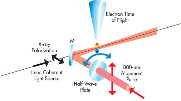 Figure 1. Schematic of a typical laser pump/x-ray probe experiment showing how the pulses are overlapped in the sample and analyzed. (In this case, the resultant data “signals” are Auger electrons whose energy is analyzed via their time of flight. Figure reprinted with permission from J.P. Cryan et al, Phys Rev Lett, 105 (2010), 083004. Copyright 2010 by the American Physical Society.
Molecular alignment applications
A particularly innovative class of experiments using these lasers involves molecular alignment. Here the ultrafast laser is used to align molecules in the lab frame. The aligned molecules are then probed by polarized x-ray pulses from the FEL (see Figure 1). A very successful example of this has been examining how Auger electrons are emitted relative to the molecular frame (principal axes).2
In Auger spectroscopy, high-energy photons or electrons remove an electron from the inner core of an atom. As shown in Figure 2, this eventually causes an outer shell (valence) electron to be ejected. In molecular systems, the kinetic energy distribution of this electron contains fine details related to the chemical bonds in the molecule. For example, a nitrogen atom bonded to a carbon atom will produce a pattern different from that of a nitrogen atom bonded to another nitrogen atom. The availability of femtosecond x-ray pulses thus offers the potential to use Auger spectroscopy to probe changing chemical conditions on the femtosecond scale; i.e., on the timescale of the bond formation during a chemical reaction.
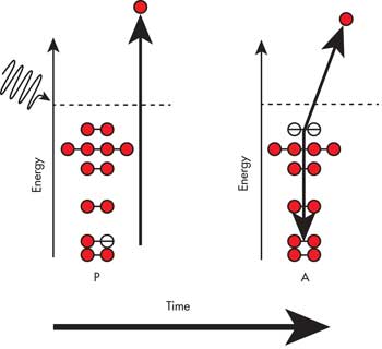
Figure 2. In Auger spectroscopy, a high-energy (e.g., x-ray) photon causes an inner-core electron to be eliminated from the target atom or molecule. A valence electron then drops down to fill the resultant orbital vacancy. This creates excess energy, which is released by ejection of another valence electron whose kinetic energy is measured by time of flight or similar means. A detailed examination of the kinetic energy of the released electrons from a sample provides important information about the local chemistry of the sample.
Scientists using the LCLS FEL recently advanced this research in two ways that enable novel measurements of molecular dynamics.2 First, they used the high intensity of the FEL pulses to remove two core electrons from nitrogen (N2) molecules (Figure 2). Second, they used an intense, 800-nm polarized pulse from a Ti:sapphire laser to drive coherent Raman interactions with the rotating N2 molecules. The Raman interaction changes the rotational quantum state of the molecule while also aligning the axis of rotation of a significant population of the N2 molecules to the polarization direction of the laser beam. As a result, the axes of the molecules under examination are no longer randomly arranged but are preferentially aligned to a laboratory-fixed direction. This direction can be switched at will simply by rotating the polarization direction of the ultrafast laser beam.
A polarized FEL pulse then drives the Auger process before any collisions or other mechanisms can destroy the alignment. The first published results from this landmark experiment show that, as expected from theory, the intensity of Auger electrons emitted using lower x-ray energies is dependent on the orientation of the molecules relative to the polarization of the x-rays (Figure 3).
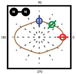
Figure 3. Angular yield of Auger electrons for 90°, 45° and 0° (blue, green, red) with respect to the molecular axis for double-core (two electrons) vacancies in prealigned nitrogen molecules. Clearly, this distribution peaks at 0° molecular orientation. Figure reproduced with permission from Physical Review Letters.
Matter in extreme conditions
High-energy-density physics is concerned with matter in states of extreme pressure and temperature, such as the conditions found in stars, the cores of large planets and nuclear fuel under inertial confinement in fusion research. Many unanswered fundamental questions remain about the physics of materials in these conditions, and ultrafast x-ray FELs now provide unique capabilities for measuring the dynamic properties of the short-lived high-energy density states. For example, whether the standard gas equation (PV = nRT) holds under these extreme conditions is still an open question. Since the 1970s, pulsed lasers have been used to drive materials into these extreme conditions by rapid heating and compression of materials, and significant extension of the range of available energy density has been enabled by amplified ultrafast lasers producing intensities above 1018 W/cm2.
The LCLS Matter in Extreme Conditions instrument is being commissioned at SLAC specifically to explore this area of physics. The instrument comprises a large target chamber, a suite of x-ray diagnostics and several laser systems, including a 50-J, few-nanosecond Nd:glass laser and a terawatt 150-mJ, 35-fs Ti:sapphire laser. Examples of the experiments proposed to take advantage of this capability include heating materials with optical lasers and using x-ray diffraction, scattering and absorption spectroscopy to probe the equation of state. Other experiments will involve creating high-pressure matter by driving a shock wave through a material with an optical laser and probing the state of the compressed lattice with x-rays. And yet others will use x-rays to rapidly heat materials before using ultrafast lasers for time-resolved interferometric measurement of the velocity of the surface of the material.
Synchronizing FEL pulses, fs lasers
Taking full advantage of the temporal resolution in experiments involving ultrafast and FEL pulses requires timing control of the variable delay between the pulses on a timescale of 30 fs or less. There are two complementary approaches to meeting this challenge. A fundamental requirement is active synchronization of the ultrafast laser to the FEL master radio-frequency oscillator. To date, however, various sources of timing jitter in the x-ray beam at LCLS are difficult to reconcile and limit this synchronization to the >100-fs (rms) level. For many experiments where phenomena on the 10-fs timescale are important, this level of jitter is not adequate and requires a second approach: measuring the relative timing between the laser and x-rays on a shot-by-shot basis. The raw data is then re-sorted, with every recorded data point re-binned according to the measured delay between the x-rays and the optical laser.
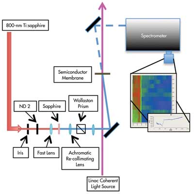
Figure 4. (a) 100 nm of continuum is generated by focusing 800-nm Ti:sapphire pulses into 1-mm-thick C-plane cut sapphire. A reference HeNe is co-propagated with the 800-nm light. The continuum spectrum is shown in the inset (b). The spectrometer limits the effective bandwidth to a 620- to 700-nm smooth region that is used in this experiment. The pulses are chirped to 3 ps with 2 in. of SF11 glass. (c) The continuum then probes the Si3N4 membrane. The x-rays change the index in the membrane, thus changing the spectral transmission of the probe (d). Two equivalent spectra are separated into two distinct stripes, as shown in the inset (e). The reference HeNe spectral line is seen on the right side of this image. Figure reproduced with permission from Optics Express.
Obviously, this requires a method of reliably measuring the temporal separation of the x-ray/ultrafast pulses on every shot. Eventually, scientists would like the ability to see the complete temporal profile of the x-ray pulse, which will require complete temporal profile retrieval methods such as frequency-resolved optical gating in the x-ray domain using a physical process that is sensitive to these x-rays and significantly faster than 30 fs. Scientists at SLAC and other FEL facilities have shown that relative timing can be measured by imparting a spectral and/or spatial chirp to the ultrafast pulse. This then reflects off – or is transmitted through – a material whose optical properties are dynamically modified by the x-ray pulse.
In one such method, a small fraction of the ultrafast laser pulse is collimated and reflected off a silicon nitride (Si3N4) membrane at a 50° angle of incidence before reaching a CCD camera.3 Using this large angle of incidence causes the time at which the pulse reaches the membrane to vary linearly across the collimated beam. The collimated x-ray pulse from the FEL passes through this membrane at normal incidence, releasing a large population of charge carriers and temporarily causing a change in the membrane’s refractive index and, hence, optical reflectivity. This geometry effectively maps various relative arrival times of the two pulses across the profile of the ultrafast beam that can be seen in the CCD camera output; the temporal overlap between the optical laser and the x-rays manifests as a step in the intensity of the reflected ultrafast beam profile. Because this method uses only a small portion of the ultrafast pulse intensity, and because more than 80 percent of the x-ray intensity passes through the membrane, sufficient x-ray flux is available for the actual experiment.
In a related study, the ultrafast laser pulse is focused into a sapphire disk to create a supercontinuum that is chirped by transmitting it through a few centimeters of glass.4 A smooth section of this continuum (620 to 700 nm) is then transmitted through a thin Si3N4 membrane. Again, this is irradiated at normal incidence by the collimated x-ray pulse, which causes a transient change in refractive index and, in this case, optical transmission. The optical pulse is then dispersed in a spectrograph to produce a time-dependent distribution of wavelength that is recorded with a CCD. A step function in the intensity plot indicates the onset of the x-ray pulse with a measured rms accuracy of <25 fs.
Ultrafast laser system requirements
LCLS is equipped with a considerable array of Ti:sapphire-based ultrafast laser oscillators and amplifiers, such as Coherent’s Vitara (oscillator) and Legend (amplifier), as well as various wavelength-extension accessories. To take full advantage of the FEL’s capabilities, the ultrafast laser should provide state-of-the-art performance and flexibility and yet be reliable and easy to use.
For example, many experiments require external timing synchronization to the FEL’s master radio-frequency system with a jitter of 100 fs or better. This simplifies data processing and eliminates the need for pulse correlation measurements in many applications. In addition, with an LCLS beam time cost of more than $30,000 per hour, 24/7 reliability and minimum maintenance are mandatory! The decision to purchase Coherent Vitara oscillators was determined partly by the fact that these next-generation flexible lasers are one-box turnkey instruments supporting hands-free operation over thousands of hours, while also delivering state-of-the-art performance specifications.
Other key laser requirements of the full amplifier system include high shot-to-shot stability, with fluctuations <0.5 percent and straightforward flexibility in switching between quite different wavelength domains from the UV through mid-IR – even producing teraherz with appropriate user setup.
Meet the authors
Alan R. Fry is deputy director of the Laser Science and Technology division at SLAC National Accelerator Laboratory in Menlo Park, Calif.; email: [email protected]. Marco Arrigoni is director of marketing at Coherent Inc. in Sunnyvale, Calif.; email: marco.arrigoni @coherent.com.
References
1. M. Först et al (December 2011). Driving magnetic order in a manganite by ultrafast lattice excitation. Phys Rev B, Vol. 84, Issue 24, 241104(R).
2. J.P. Cryan et al (August 2010). Auger electron angular distribution of double core-hole states in the molecular reference frame. Phys Rev Lett, Vol. 105, Issue 8, p. 083004.
3. M. Beye et al (March 2012). X-ray pulse preserving single-shot optical cross-correlation method for improved experimental temporal resolution. Appl Phys Lett, Vol. 100, Issue 12, p. 121108.
4. M.R. Bionta et al (October 2011). Spectral encoding of x-ray/optical relative delay. Opt Exp, Vol. 19, No. 22, p. 21855.