Fluorescence lifetime imaging microscopy derives its information from the decay rates of individual fluorophores. It is opening up understanding of cell metabolism, inner cell reactions and cell death.
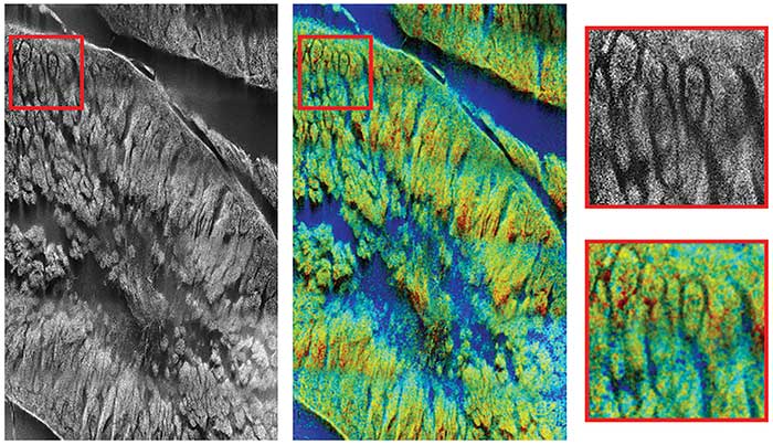 Fluorescence lifetime imaging microscopy (FLIM) enables researchers in the life sciences to get information from live specimens about interactions on the molecular scale. The technique captures the differences in the excited state decay rate from a fluorescent sample, rather than relying on the concentration of a fluorophore. Since imaging does not derive from the intensity of a signal, the technique lessens the impact of photon scattering in thick layers of sample and is generally considered more robust than intensity-based methods. This can open doors for both researchers and clinicians.
To gain a deeper understanding of what FLIM has to offer, we asked a panel of academic researchers and industry experts to talk about the strengths, weaknesses and future of this powerful imaging tool. FLIM has a range of applications — from tracking the effects of drugs in real time and guiding measurements during surgery to detecting energy transfer from FRET (Förster or fluorescence resonance energy transfer).
FRET FLIM is the change in fluorescence lifetimes due to the presence of energy transfer from FRET. In the presence of FRET, you see shortened donor molecule lifetimes as energy is donated to the acceptor. This provides a quantitative method of verifying that FRET is occurring. FRET FLIM is powerful because it provides data independent of dye or protein concentration, photobleaching and excitation light intensity — that is, laser power. Quantitative FLIM studies are sometimes done to record changes in the intracellular environment around the fluorescent molecules of interest. If an environment becomes more acidic, or if the researcher fixes the cell, the lifetimes change. FLIM allows the scientist to watch the physiology of the cell in terms of pH or redox state changes or other perturbations of the cell.
Fluorescence lifetime imaging microscopy (FLIM) enables researchers in the life sciences to get information from live specimens about interactions on the molecular scale. The technique captures the differences in the excited state decay rate from a fluorescent sample, rather than relying on the concentration of a fluorophore. Since imaging does not derive from the intensity of a signal, the technique lessens the impact of photon scattering in thick layers of sample and is generally considered more robust than intensity-based methods. This can open doors for both researchers and clinicians.
To gain a deeper understanding of what FLIM has to offer, we asked a panel of academic researchers and industry experts to talk about the strengths, weaknesses and future of this powerful imaging tool. FLIM has a range of applications — from tracking the effects of drugs in real time and guiding measurements during surgery to detecting energy transfer from FRET (Förster or fluorescence resonance energy transfer).
FRET FLIM is the change in fluorescence lifetimes due to the presence of energy transfer from FRET. In the presence of FRET, you see shortened donor molecule lifetimes as energy is donated to the acceptor. This provides a quantitative method of verifying that FRET is occurring. FRET FLIM is powerful because it provides data independent of dye or protein concentration, photobleaching and excitation light intensity — that is, laser power. Quantitative FLIM studies are sometimes done to record changes in the intracellular environment around the fluorescent molecules of interest. If an environment becomes more acidic, or if the researcher fixes the cell, the lifetimes change. FLIM allows the scientist to watch the physiology of the cell in terms of pH or redox state changes or other perturbations of the cell.
Member Exclusive: To read the complete article, please Login or Register