FTIR spectroscopy has become a dominant tool for examining biological samples in the study of neurological and other diseases, from Alzheimer’s to cancer.
DANIEL MANN, CARSTEN KÖTTING, AND KLAUS GERWERT, RUHR-UNIVERSITÄT BOCHUM
Almost every molecule in the universe interacts with IR radiation, which excites vibrations in the molecule, rendering Fourier transform IR (FTIR) spectroscopy a universal method to investigate biological systems.
In contrast to many popular biophysical techniques — such as fluorescence spectroscopy, x-ray crystallography, or cryoelectron microscopy — samples in FTIR spectroscopy can be investigated label-free at room temperature in solution. Most structural methods yield only static snapshots of a dynamic reaction, whereas FTIR spectroscopy enables time-resolved investigation at near-native conditions, without labels that can severely interfere with the sample or the reaction.
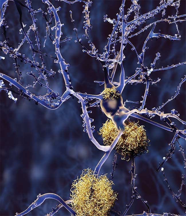
FTIR spectroscopy is bringing scientists deeper inside
biological samples such as neurons (shown here invaded by amyloid plaques), allowing more extensive research of diseases such as Alzheimer’s.
In most instances, IR spectroscopy is performed in transmission or via reflection at surfaces. Most commonly, broadband IR light is obtained from a thermal source. Spectral resolution is gained by interferometry, where a moving mirror within the Michelson interferometer leads to interference, depending on the displacement of the mirror. Fourier transformation yields the single-channel IR spectrum (Figure 1).
An absorbance spectrum depicts specific absorbance peaks of the sample relative to a reference and is calculated according to the Beer-Lambert law. The absorbance occurs in a linear relationship with the concentration of an absorbing species. Hence, FTIR spectroscopy enables quantitative analysis; for example, the stoichiometry of a protein and a ligand during a reaction can be determined.
FTIR spectroscopy has significant advantages over conventional spectroscopy using dispersive elements, resulting in a very good signal-to-noise ratio. Recorded IR spectra are very precise. In the case of phosphate molecules, an IR shift of 1 cm-1 corresponds to a P-O bond length alteration of 0.001 nm. Complex information — such as bond lengths and angles, aggregate state, secondary structure of proteins, solvation (an interaction of a solute with the solvent), and environment, including hydrogen bonds and much more — is contained in recorded IR spectra. This high resolution is essential for determining catalytic mechanisms, as one single hydrogen bond can accelerate a reaction by several orders of magnitude.
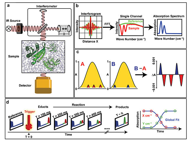
Figure 1. Principle of FTIR spectroscopy. IR radiation is modulated in a Michelson interferometer (consisting of a beamsplitter as well as fixed and movable mirrors), which creates multiwavelength interference (a). Recorded interferograms of reference and sample are Fourier transformed, and an absorption spectrum is calculated (b). As almost every molecule in the sample contributes to the IR spectrum, changes during a reaction are small compared to the overall signal (c). Difference spectroscopy eliminates the background (yellow) and makes small changes (red and blue) visible. Time-resolved FTIR difference spectroscopy requires a reaction trigger and enables investigation of biological reactions with high spatio-temporal resolution (d). FFT: fast Fourier transform. Courtesy of Ruhr-Universität Bochum.
Ubiquitous absorption of IR radiation leads to the contribution of all reactants and products of the reactions — proteins, lipids, ligands, and the solvent — to the IR spectrum. Water (essential for the majority of biological processes) contributes greatly to IR spectra, and constant solvation dynamics of water, which interacts with the sample, superimposes measured IR spectra.
One challenge in IR spectroscopy is to resolve the tiny signals that are caused, for example, by an enzymatic reaction behind this large background. Difference spectroscopy removes the static background and resolves small changes that occur, such as in enzymatic reactions (Figure 1c). The targeted introduction of tiny changes alters only a small number of bands within the recorded IR spectra, which can then be assigned to specific elements of the system. Once obtained, the high-resolution information is still encoded into a spectrum that is difficult to interpret, and the origin of each band has to be assigned for a deeper understanding. This can be achieved via isotopic labeling, point mutations, or hydrogen-deuterium (HD) exchange. Another comprehensive approach is to calculate the experimental IR spectrum from biomolecular simulations.
For time-resolved FTIR, sharp triggers are required for a defined start of the reaction. UV/VIS light is an ideal reaction trigger that can be applied externally without causing too much perturbation in the system, as would occur with techniques such as rapid mixing. The field of time-resolved FTIR spectroscopy was therefore developed with photoactivatable proteins such as bacteriorhodopsin1. Application of photoactivatable compounds — including pHPcgATP, NPEcgGTP, photocaged Mg2+, or photocaged NADP — have opened the field for proteins that are intrinsically inert to UV/VIS radiation2. FTIR spectroscopy has been used extensively to investigate molecular mechanisms in GTPase and ATPase proteins.
GTPase proteins are molecular switches that regulate a plethora of physiological processes in the body, including vision, scent, and blood pressure regulation, and are key to severe diseases such as cancer. ATPase proteins are equally important, although they are more often associated with transport processes, energy generation, and storage, and are fundamental to muscle movement.
This has enabled elucidation from mechanisms on the level of macromolecular organization of proteins on lipid membranes, to the secondary structure of individual proteins, to the mechanism of single amino acids3, down to water molecules4 and single hydrogen atoms5 (Figure 2).
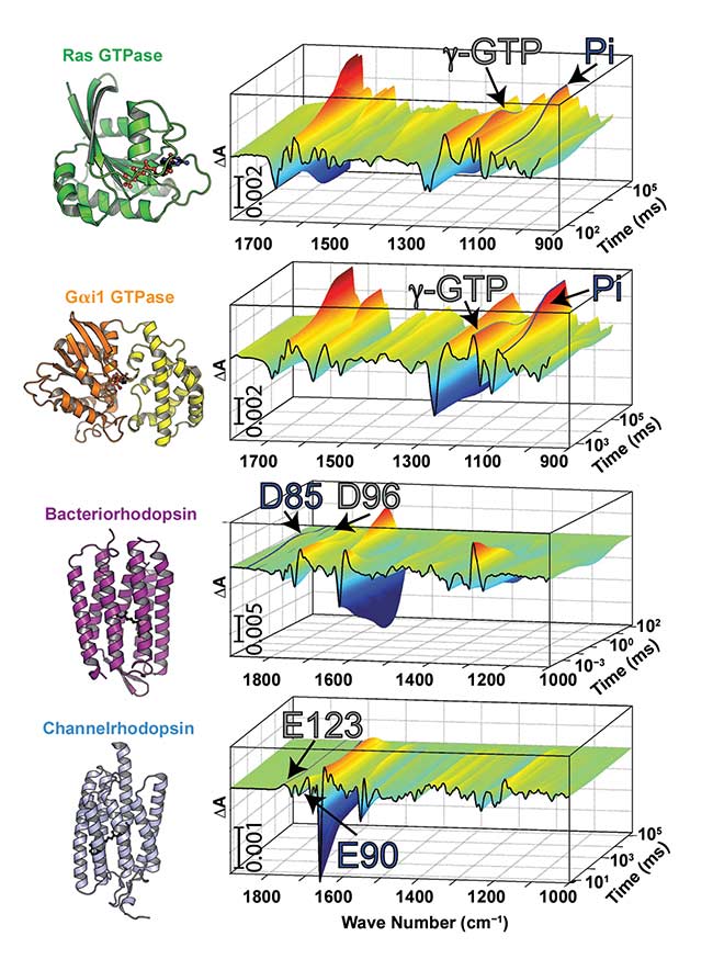
Figure 2. Time-resolved FTIR spectra of the GTP hydrolysis reaction in the small GTPase Ras (PDB-ID 1QRA) with the trigger NPEcgGTP of the GTP hydrolysis
reaction in the heterotrimeric GTPase Gai1, using the trigger pHPcgGTP (PDB-ID 1GIA) of the photocycle of the proton pump bacteriorhodopsin (PDB-ID 1QHJ), and the photocycle of the optogenetic tool channelrhodopsin-2 (PDB-ID 6EID). Bands for the third phosphate group γ-GTP and the cleaved GTPase product Pi are labeled for Ras and Gai1. Acidic amino acids that undergo protonation change during the photocycles of bacteriorhodopsin and channelrhodopsin-2 are further indicated. Courtesy of Ruhr-Universität Bochum.
Temporal resolution in FTIR spectroscopy is limited by the velocity of the moving mirror within the Michelson interferometer and is typically in the millisecond time range. However, recent developments in both step-scan FTIR spectroscopy and IR light sources further increase temporal resolution to the femtosecond timescale.
Quantum cascade lasers (QCLs) in the IR range possess tuneable wavelengths, rendering the whole interferometer redundant for proteins that have a reaction cycle, that is, that can be excited several times. In addition, QCLs are much brighter than conventional blackbody radiators, which further increases the signal-to-noise ratio. Utilization of QCLs also enables the advantage of frequency combs that can be used to simultaneously record a broad spectral range within several microseconds6. This technique will hopefully enable measurements of single-shot kinetics of noncyclic reactions, such as the GTP hydrolysis reaction using QCL-IR spectroscopy, in the future.
FTIRspectroscopy coupling
IR spectra can be directly calculated from biomolecular simulations. For the majority of cases, only costly quantum mechanical (QM) simulations that explicitly include electrons are accurate enough to precisely reproduce the experiment. However, QM simulations are computationally very expensive and restricted to system sizes of only hundreds of atoms. Protein systems are usually much bigger.
Recent advances in both computational technology and hybrid QM/MM (molecular mechanics) simulations, which embed a QM region such as an active protein center in an environment that is treated via classical MM, lead to successful reproduction of FTIR experiments (Figure 3). This technique is very precise and enables highly accurate elucidation of several protein reactions. Constant improvements in modern computer systems regularly enable faster and more accurate calculations.
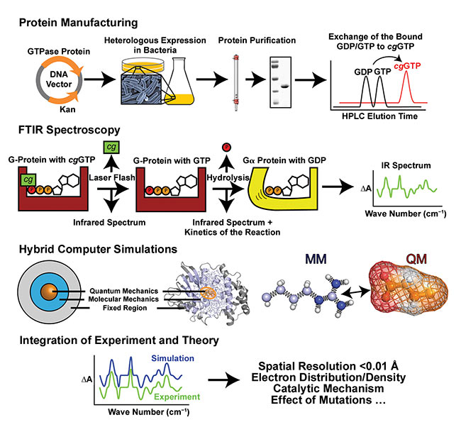
Figure 3. Investigation of a GTPase protein using IR spectroscopy. After protein production and purification, the bound nucleotide is exchanged to photocaged GTP. Using laser light as a trigger, the time-resolved FTIR experiment is performed; assignment of the obtained spectra can be performed using biomolecular simulations. Quantum mechanical (QM) simulations are required for the accurate calculation of IR spectra. However, QM simulations are computationally very expensive and restricted to systems sized only hundreds of atoms. Protein-ligand systems are usually much bigger. Hybrid QM/MM (molecular mechanics) computer simulations enable calculation of vibronic and electronic properties of protein-active centers that are treated quantum mechanically and embedded into an environment that is treated via MM. If calculated IR spectra match experimental spectra, enzymatic reactions can be followed with very high spatio-temporal resolution, and catalytic mechanisms can be elucidated. Courtesy of Ruhr-Universität Bochum.
If theoretical and experimental IR spectra match, the experiment can be translated to a 3D movie of the reaction with precise information of every atom and even its charge distribution3. High spatiotemporal resolution and spectral accuracy of time-resolved FTIR spectroscopy, together with the high accuracy of biomolecular simulations, renders integration of both methods as the perfect toolbox for investigation of biological processes.
A diagnostic tool
The power of IR spectroscopy, along with its increasing capabilities enabled by QCLs, has motivated recent application for diagnostics of neurodegenerative diseases7, diabetes8, and cancer9,10. The position of the C=O stretching vibration in the protein backbone (amide I band) is sensitive to the fold of the protein. This can be exploited to investigate diseases that are presumably caused by misfolded proteins, such as in Alzheimer’s disease7. Amyloid beta protein can be captured by custom antibodies in an attenuated total reflection (ATR)-FTIR system, and the position of the amide I band elucidates whether amyloid beta is present in the form of helices or beta sheets.
A recent study by Nabers et al. was able to show that beta-sheet formation leads to development of Alzheimer’s, even years before the disease outbreak7, opening a vision on early detection. Moreover, IR microscopes are now powerful tools that can be used to perform label-free pathology9 (Figure 4). Classification of the spatially resolved IR fingerprint enables distinction of several tissue types, even if the investigated cells have developed cancer9. QCLs have accelerated this process by a factor that allows for both label-free clinical pathology within the same time range and accuracy as state-of-the-art, gold-standard pathology. However, large amounts of acquired data raise the need for accurate and high-throughput bioinformatics now and into the future. The vision is to create a noninvasive tool that can distinguish malignant and benign tissue during a coloscopy, for example.
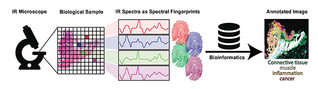
Figure 4. Label-free pathology using an IR microscope is able to annotate biological samples, such as human tissue, using classification of IR fingerprints. Powerful bioinformatics is required to deal with amounts of data. Courtesy of Ruhr-Universität Bochum.
Integration of experiment, theory, and bioinformatics, together with further improvements in stability and affordability of QCLs, has increased signal-to-noise ratio in single-shot kinetics. Integration of other biophysical techniques, such as fluorescence spectroscopy, in addition to FTIR, will be challenges to tackle in the future of IR spectroscopy.
Meet the authors
Daniel Mann is a postdoctoral research associate at the University of Sheffield in England. He has a master’s degree in structural biology from Ruhr-Universität Bochum in Germany, where, during his Ph.D. program, he studied and applied time-resolved FTIR spectroscopy and QM/MM simulations to investigate
catalytic mechanisms of small and heterotrimeric GTPases; email: [email protected].
Carsten Kötting is a group leader in the Department of Biophysics at Ruhr-Universität Bochum in Germany. He received his Ph.D. in physical organic chemistry from Ruhr-
Universität. He is a former Feodor Lynen Fellow of the Zewail group at Caltech and is currently researching details of the reaction mechanisms of GTPases, and development of chemically modified surfaces for protein immobilization on ATR crystals; email: [email protected].
Klaus Gerwert is a professor and chair of the Department of Biophysics at Ruhr-Universität Bochum. He received his Ph.D. in biophysical chemistry from the University of Freiburg in Germany. He is a former fellow of the Max Planck Society and served as director of the Max Planck Partner Institute for Computational Biology (PICB) in Shanghai. In 2010, he founded the European Protein Research Unit Ruhr within Europe (PURE) with clinicians. He is the founding director of ProDi, a federal/state-financed research center for molecular protein diagnostics; email:
[email protected].
References
1. K. Gerwert et al. (1990). Simultaneous monitoring of light-induced changes in protein side-group protonation, chromophore isomerization, and backbone motion of bacteriorhodopsin by time-resolved Fourier-transform IR spectroscopy. Proc Natl Acad Sci USA, Vol. 87, Issue 24, pp. 9774-9778.
2. C. Kötting et al. (2006). A phosphoryl transfer intermediate in the GTPase reaction of Ras in complex with its GTPase-activating protein. Proc Natl Acad Sci USA, Vol. 103, Issue 38, pp. 13911-13916.
3. D. Mann et al. (2016). Mechanism of the intrinsic arginine finger in heterotrimeric G proteins. Proc Natl Acad Sci USA, Vol. 113, Issue 50, pp. E8041-E8050.
4. F. Garczarek et al. (2006). Functional waters in intraprotein proton transfer monitored by FTIR difference spectroscopy. Nature, Vol. 439, pp. 109-112.
5. D. Mann et al. (2018). The protonation states of GTP and GppNHp in Ras proteins. J Biol Chem, Vol. 293, pp. 3871-3879.
6. J.L. Klocke et al. (2018). Single-shot sub-microsecond mid-IR spectroscopy on protein reactions with quantum cascade laser frequency combs. Anal Chem, Vol. 90, Issue 17, pp. 10494-10500.
7. A. Nabers et al. (2018). Amyloid blood biomarker detects Alzheimer’s disease. EMBO Mol Med, Vol. 10, Issue 190.
8. M.A. Pleitez et al. (2013). Windowless ultrasound photoacoustic cell for in vivo mid-IR spectroscopy of human epidermis: low interference by changes of air pressure, temperature, and humidity caused by skin contact opens the possibility for a non-invasive monitoring of glucose in the interstitial fluid. Rev Sci Instrum, Vol. 84, Issue 8, 084901.
9. A. Kallenbach-Thieltges et al. (2013). Immunohistochemistry, histopathology and IR spectral histopathology of colon cancer tissue sections. J Biophotonics, Vol. 6,
Issue 1, pp. 88-100.
10. P. Lasch et al. (2004). Imaging of colorectal adenocarcinoma using FT-IR microspectroscopy and cluster analysis. Biochim Biophys Acta Mol Basis Dis, Vol. 1688, Issue 2, pp. 176-186.