
Fiber Optic Probes Help Customize Spectroscopic Diagnosis
When a sample is illuminated at a specific wavelength, the emitted fluorescence reveals details within a sample’s composition, speeding disease identification.
AMANDINE DEBLOUDTS, SEDI-ATI FIBRES OPTIQUES
Diffuse reflection spectroscopy and fluorescence spectroscopy are having a major effect in medicine and the life sciences, with expanding application in physics, chemistry, biology, and medicine. And fiber optic-based probes are becoming an essential and versatile solution for collecting the necessary spectroscopic measurements for analysis. This data ultimately informs both in vivo and in vitro analysis and diagnosis to detect cancer cells or the presence of specific diseases and may in some instances render traditional biopsies unnecessary.
Blood analysis
Fiber optic probes are ideal for performing fluorescence spectroscopy in conjunction with in vitro blood analysis. They allow a scientist or clinician to illuminate a sample of blood at a defined wavelength and to collect the fluorescence emitted as a reaction to the excitation caused by the incoming light. The emitted fluorescence is directly related to the composition of the target, which allows the scientist or clinician to identify a particular disease or condition.
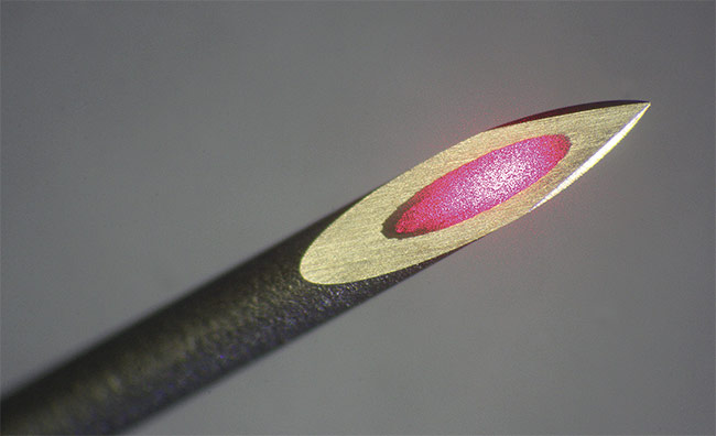
Fiber optics contained within a hypodermic needle can deliver targeted excitation of fluorescence to a tumor. Courtesy of SEDI-ATI.
Fiber optic probes, such as those utilizing bifurcated bundles with multiple branches, can be used to analyze several samples of blood at once, with the aim of providing a rapid and accurate signature-based measurement for major chronic diseases. Such technology is already used routinely in clinical laboratories for the screening of protein abnormalities in the blood of patients.
In the case of bundled probes, the fiber assemblies can have an “octopus” design, consisting of several fibers arranged in a linear, circular, or matrix fashion at one end of the octopus and a fanout of several individual fibers at the other end. Each fiber optic leg brings a specific wavelength paired with a particular reagent. The stimulated reagent produces fluorescence, which is detected by an additional fiber. Then, there are as many samples that can be tested simultaneously as there are channels (legs) available on the octopus.
Such assemblies can include fibers with core diameters ranging from 50 to 1000 µm and lengths of several tens of meters. Of course, when it is installed in an instrument, there is no need for meters of fibers. There are some cases, however, when the electronics must be deported from the sample because the measurement is completed in a room or chamber with a particular atmosphere (vacuum, temperature, cryogeny, radiation, etc.) that is not safe for people or instruments. In other cases, the sources and detectors are in one room, while the fibers must serve another.
The basics of fiber optics
Fiber optics is a versatile, low-cost solution that offers numerous advantages for diffuse reflectance spectroscopy in comparison to other types of instrumentation. Fiber optics enable a wide wavelength range between 185 and 2.5 µm due to solarization-resistant fibers and infrared fibers. It allows considerable space and weight savings by offsetting both the light source and the detector, either a monochromator or spectrometer. Additionally, users can isolate and stabilize electronic systems, including laser sources and spectrometers, that are sensitive to environmental parameters such as radiation or hot and cold temperatures.
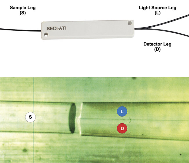
Multimode couplers can be incorporated into a fiber optic probe to provide direction and spectral
resolution, while enabling the use of broadband light sources. Courtesy of SEDI-ATI.
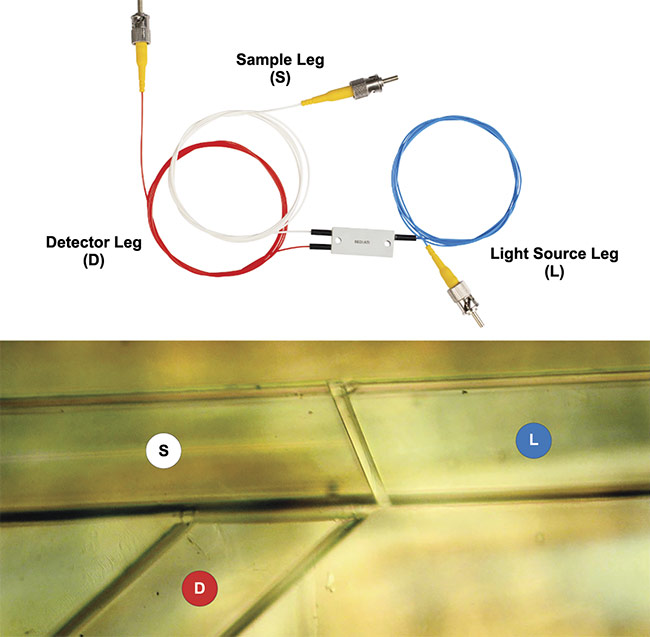
Wavelength division multiplexer probes allow for a variety of fibers to be used along with a dichroic filter to deliver excitation and collect emission from a selected wavelength. Courtesy of SEDI-ATI.
There are three different fiber-based optical probe technologies that are relevant to diffuse reflectance spectroscopy measurements. Each has its own advantage. To facilitate in vivo or in situ diagnosis, all three of these probe types can be combined with a needle tip.
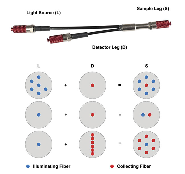
Reflection probe fiber bundles
These bundles generally comprise optical fibers in a round or linear configuration combined in a ferrule or SMA905 connector to match the entrance slit of most spectrometers. This configuration allows the use of large-core step-index multimode fibers of up to 1000 µm. The bundles can also contain a large number of fibers, which could include several collecting fibers around a larger illumination fiber, or several illumination fibers around a larger collecting fiber. In addition, it allows different detection geometries. Lastly, fibers can be arranged randomly or coherently, according to the needs of the application. For example, for blood analysis and diagnosis, the fibers must be coherently arranged for traceability, meaning the exact position of each fiber is known from one end of the probe to the other, which avoids any confusion in the results.
Multimode coupler probes
These probes incorporate a 1 × 2 multimode coupler that offers the great advantage of high directivity, which minimizes the light that can pass directly from the injection port to the output port. This solution, based on GI50 or GI62.5 gradient-index fibers, offers better spectral resolution than bifurcated fiber bundles. Finally, it is mode-insensitive and achromatic over the 400- to 1625-nm wavelength range, enabling the use of broadband light sources that can maximize fluorescence potential from the UV to the NIR without affecting the coupling ratio. Their high directivity prevents noise from being a factor, which would be an issue for fused couplers.
Multiplexer probes
The heart of this device is a dichroic filter that can be custom-made for use in UV, VIS, and IR spectra, enabling a wide range of disease detection. It enables the selection of the main wavelengths of interest, for both guiding incident light to the target and directing collected light to the detector. This makes it ideal for fluorescence detection because it separates the targeted signal from other wavelengths, thus reducing the noise, because bundles intrinsically catch all the light that can be transported by the fiber. Couplers also catch noise but less so than bundles — one collecting fiber of a 50-µm core diameter versus one to six large-core collecting fibers with core diameters that can reach 1000 µm each. The lower the diameter of the fiber core, the less light that can be caught in free space.
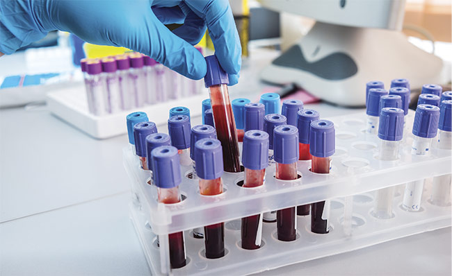
The need to test multiple blood samples at once has spawned innovation in fiber optic probes. Courtesy of Romaset — stock.adobe.com.
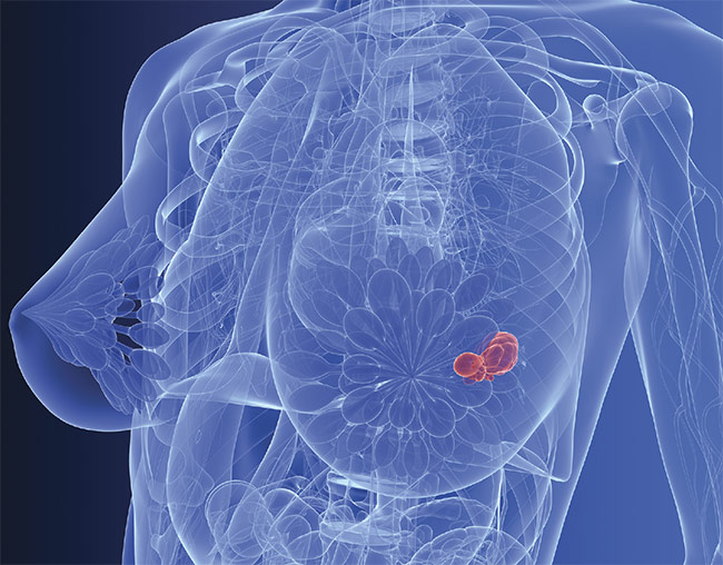
Breast cancer tumors have traditionally been the subject of extensive biopsy procedures, though new fiber optic technology could simplify the procedure. Courtesy of SciePro — stock.adobe.com.
These benefits have been used in a variety of forms that could prove to have a myriad of clinical benefits, such as in diagnostics, in which current medical practice requires a number of steps for alternative forms of analysis.
Breast cancer detection
Another medical application in which fiber optic probes can be used is the early detection of breast cancer, since detection at an early stage of development can lead to more effective treatment. Current protocol dictates when a suspicious mass is detected by mammography, the patient must undergo tests to determine whether it is a benign or malignant tumor. But a mammogram or ultrasound cannot definitively show whether cancer cells are present in the mass.
In practice, it is therefore necessary to perform a percutaneous tissue sampling (biopsy) to analyze the cells in situ. This invasive procedure is often painful for the patient since it involves making an incision of a few millimeters to introduce a large needle up to the suspect lesion. Several samples are taken using the needle to cut tissue fragments. The procedure can last up to an hour under local anesthesia and can cause lingering discomfort, especially when it is a macro biopsy. The samples from the biopsy are then analyzed by a specialized laboratory, and the result is not known until several days later.
Fiber bundles of varying sizes can be grouped in various geometries, depending on the application. Courtesy of SEDI-ATI.
It should be noted that experts have argued in the American Journal of Surgical Pathology1 that this routine examination should not be carried out routinely, given that 80% of biopsies have proved to be negative for cancerous tissue. To avoid this painful — and often emotional — experience for women who have had benign abnormalities identified, an innovative medical probe made of an optical fiber inside of a hypodermic needle can instead be used to perform in vivo, real-time investigation with fluorescence spectroscopy. Not only is this method more comfortable for patients but it is substantially faster than collecting and analyzing biopsy results, and potentially reassures patients far sooner in their clinical visits. Many biopsies require between 45 and 60 minutes, whereas the fiber-in-a-needle procedure could take about 15 minutes.
How the probe works
The aforementioned medical probe, developed by SEDI-ATI in conjunction with a startup called NODEA MEDICAL a number of years ago, consists of a small hypodermic needle that provides excellent tissue penetration. The hypodermic needle is 0.5 mm, similar to a needle used to take blood samples through the insertion of an optical fiber with a 200- to 600-µm core diameter. The profile of the needle, similar to that of vaccine needles, allows it to pass through the skin without a prior incision. Under ultrasound guidance, the probe is introduced to the suspected lesion. Then, it illuminates the cells with laser light.
Without even injecting a marker product or fluorochrome, this technique causes the cells to fluoresce; in other words, they emit their own light in response to the excitation caused by the incident light. This light is captured and guided to a detector by the same optical fiber. By observing how the illuminated tissue scatters the light, the specialist can immediately confirm and refine his or her diagnosis of suspicious lesions. While this technique is not intended to eliminate biopsies altogether, it could very well serve to significantly reduce the number of unnecessary biopsies in medical practice.
Meet the author
Amandine Debloudts joined SEDI-ATI Fibres Optiques in June 2018 and became the market communications manager. Following technical studies in optronics, she started as an application engineer and a technical sales engineer from 2005 to 2012 within SME SDS High Voltage, and then at AMS Technologies, a pan-European provider of custom products and solutions with competencies across photonics, thermal management, and power technologies. She has also worked in marketing and communications positions at 3S Photonics, Innov-Plus, and IncubAlliance; email: [email protected].
References
1. C.J. VandenBussche et al. (2016). Reflex estrogen receptor (ER) and progesterone receptor (PR) analysis of ductal carcinoma in situ (DCIS) in breast needle core biopsy specimens: an unnecessary exercise that costs the United States $35 million/y. Am J Surg Path, Vol. 40, No. 8, pp. 1090-1099.
/Buyers_Guide/SEDI-ATI_Fibres_Optiques/c13332
Published: September 2023
Glossary
- fluorescence spectroscopy
- The spectroscopic study of radiation emitted by the process of fluorescence.
- fluorescence
- Fluorescence is a type of luminescence, which is the emission of light by a substance that has absorbed light or other electromagnetic radiation. Specifically, fluorescence involves the absorption of light at one wavelength and the subsequent re-emission of light at a longer wavelength. The emitted light occurs almost instantaneously and ceases when the excitation light source is removed.
Key characteristics of fluorescence include:
Excitation and emission wavelengths: Fluorescent materials...
Featuresfiber optic probesfluorescence spectroscopydiffuse reflection spectroscopyBlood Analysisbifurcated bundlesDisease Detectionbreast cancerbiopsySedi-ATIreflection probe fiber bundlesmultimode couplerfluorescence