
‘Firefly’ Imaging Technique Sheds Light on Molecular Forces
Using tools made of luminescent DNA, researchers at Emory University have visualized the mechanical forces of cells at the molecular level. The researchers demonstrated the technique on human blood platelets.
“Normally, an optical microscope cannot produce images that resolve objects smaller than the length of a lightwave, which is about 500 nm,” said Khalid Salaita, senior author of the study. “We found a way to leverage recent advances in optical imaging along with our molecular DNA sensors to capture forces at 25 nm. That resolution is akin to being on the moon and seeing the ripples caused by raindrops hitting the surface of a lake on Earth.”
The researchers turned strands of synthetic DNA into molecular tension probes that contain hidden pockets, which they then attached to receptors on a cell’s surface. Free-floating pieces of fluorescently tagged DNA served as imagers. As the unanchored pieces of DNA whizzed about, they created streaks of light in microscopy videos.
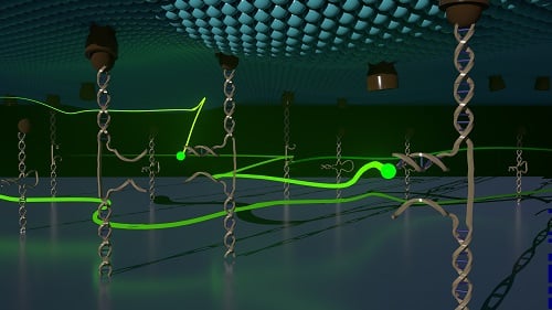
The synthetic DNA probes (brown) are anchored to a cell surface (teal). The free-floating DNA imagers are represented in fluorescent green. Courtesy of Pushkar Shinde.
When the cell in question applies force at a particular receptor site, the attached probes stretch out, causing their hidden pockets to open and release tendrils of DNA stored inside. The free-floating pieces of DNA are engineered to dock onto these DNA tendrils. When the free-floating pieces dock, they are briefly demobilized, showing up as still points of light on video.
Hours of microscopy video are taken of the process and later sped up to show how the points of light change over time, providing the molecular-level view of the mechanical forces of the cell.
The researchers used a firefly analogy to describe the process.
“Imagine you’re in a field on a moonless night and there is a tree that you can’t see because it’s pitch-black out,” said co-first author Joshua Brockman, now a postdoctoral fellow at Harvard. “For some reason, fireflies really like that tree. As they land on all the branches and along the trunk of the tree, you could slowly build up an image of the outline of the tree. And if you were really patient, you could even detect the branches of the tree waving in the wind by recording how the fireflies change their landing spots over time.”
“It’s extremely challenging to image the forces of a living cell at a high resolution,” said Su, who graduated from Emory’s Department of Chemistry and is now a postdoctoral fellow in the Salaita lab. “A big advantage of our technique is that it doesn’t interfere with the normal behavior or health of a cell.”
Another advantage is that DNA bases of A, G, T, and C, which naturally bind to one another in specific ways, can be engineered within the probe-and-imaging system to control specificity and map multiple forces at one time within a cell.
“Ultimately, we may be able to link various mechanical activities of a cell to specific proteins or to other parts of cellular machinery,” Brockman said. “That may allow us to determine how to alter the cell to change and control its forces.”
By using the technique to image and map the mechanical forces of platelets, the cells that control blood clotting at the site of a wound, the researchers discovered that platelets have a concentrated core of mechanical tension and a thin rim that continuously contracts.
“We couldn’t see this pattern before but now we have a crisp image of it,” Salaita said. “How do these mechanical forces control thrombosis and coagulation? We’d like to study them more to see if they could serve as a way to predict a clotting disorder.”
The research was published in Nature Methods (www.doi.org/10.1038/s41592-020-0929-2).
Published: September 2020