
Fluorescence Imaging Progressing from Cells to Tissue
Tissues and even whole animals are not as easily captured with fluorescence imaging as are
cells, but recent research and technological developments could change that.
Fluorescence imaging has become one of the most powerful tools for studying cellular processes. Today,
with an ever-expanding array of available fluorochromes to tag cells of interest,
scientists can identify cells and submicroscopic cellular components with high specificity
amid nonfluorescing material.
Imaging tissue is another story, however. A great challenge faces
imaging specialists, but progress is being made, and more is predicted.
The application of an array of fluorochromes (also known as fluorophores)
has made it possible for fluorescence microscopes to reveal the presence of a single
molecule. And the use of multiple fluorescence labeling makes it possible for various
probes to identify several target molecules simultaneously.
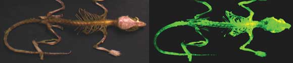
The whole skeleton of this mouse appears normal under bright-field
light (a), but it glows under fluorescence excitation with blue light (b). Courtesy
of AntiCancer Inc.
Although the fluorescence microscope cannot provide spatial resolution
below the diffraction limit of specific specimen features, detection of fluorescing
molecules below such limits is possible.
Modern fluorescence microscopes combine the power of high-performance
optical components with computerized control of the instrument and digital image
acquisition.
“At present, optical image formation is only the first step
toward data analysis,” said Allison Forlenza, assistant product and marketing
manager at Nikon Instruments Inc. in Tokyo. “Microscopy now depends heavily
on electronic imaging to rapidly acquire information at low-light levels or at visually
undetectable wavelengths.”
Computerized control of focus, stage position, optical components,
shutters, filters and detectors is in widespread use. The increasing application
of electro-optics in fluorescence microscopy has led to the development of optical
tweezers that can manipulate subcellular structures or particles, to the imaging
of single molecules and to a wide range of sophisticated spectroscopic applications.
“There are a much wider range of confocal microscopes available,
many of which are now spectral confocal, and the price range of them has dropped
considerably, with some in the $99,000 range,” said James R. Mansfield, director
of tissue analysis applications at Caliper Life Sciences Inc. in Hopkinton, Mass.
“In addition, live-cell imaging methods, environmental chambers and the ability
to track the growth of individual cells in three dimensions in embryos and cell
cultures are all growing rapidly.”
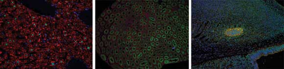
These images were taken using Nuance, a multispectral imaging system that enables imaging
of multiple molecular markers in tissue sections for fluorescence and bright field. Images courtesy of Pavol Cekan, Rockefeller University.
Naturally fluorescent proteins
A simple description of fluorescence imaging can be divided into
three parts: excitation, emission and imaging. Fluorochromes are molecules that,
when excited by a photon, will emit a photon of longer wavelength (toward the red
end of the spectrum) than that which it absorbed. This process of absorption and
re-emission is known as fluorescence.
Fluorochromes attach themselves to visible or subvisible structures
and are often highly specific in their attachment targeting. Such molecules usually
have a significant quantum yield (ratio of photon absorption to emission).
The discovery and use of naturally fluorescent proteins, which
can be used to label specific cells, has revolutionized biology by enabling the
formerly invisible to be seen clearly.
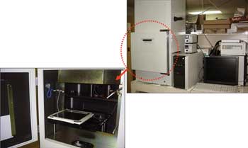
The OV100 Olympus Small Animal Imaging System offers variable magnification for imaging mice from macro to subcellular regimes. Courtesy of AntiCancer Inc.
One example is taking place at AntiCancer Inc. in San Diego. Robert
Hoffman, president and CEO, is using green fluorescent protein and red fluorescent
protein to image cancer cells in real time in live mice. Such proteins enable scientists
to visualize important aspects of cancer in living animals, including tumor cell
mobility, invasion, metastasis and angiogenesis.
“AntiCancer is the pioneer of in vivo imaging with GFP,”
Hoffman said. “This breakthrough has revolutionized imaging in small animals.
Due to our research, single cells can be followed in real time in mice, even at
the subcellular level.”
These multicolored proteins have allowed the color-coding of cancer
cells growing in vivo and enabled the distinction of host from tumor with single-cell
resolution. Hoffman and his colleagues also have used the technique to image stem
cells and bacteria in real time in live mice.
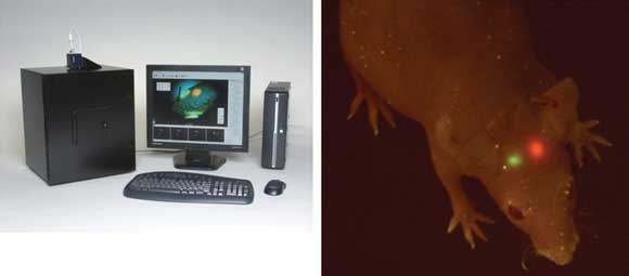
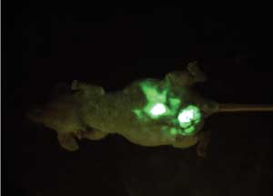
Whole-body image of GFP and RFP (red fluorescent protein) human tumors
implanted in the brain of a mouse, imaged with the Indec Systems FluorVivo imaging
system. Courtesy of AntiCancer Inc.
The company currently is developing fluorescence-guided surgery
with cancers that have been labeled with GFP and is looking to determine the efficacy
of cancer therapeutics.
“We are also developing tumor-targeting bacteria to cure
the tumors,” Hoffman said. “We have developed special mutants of salmonella
to selectively target tumors. We have labeled the bacteria with GFP to follow them
as they attack tumors.”
Tissue imaging remains a challenge
Although fluorescence imaging of cells has progressed by leaps
and bounds, when it comes to tissue imaging, the technique is not as effective.
Fluorescence imaging of tissues presents a number of challenges that are not present
in live- or fixed-cell samples and that require a different approach to obtain optimal
results.
“In cell-imaging applications, there is little to no interfering
autofluorescence signal, so highly sensitive cameras and PMT [photomultiplier tube]
detectors can be used to great effect to obtain excellent signal-to-noise ratios,”
Caliper’s Mansfield said. “For tissue imaging, however, the interfering
autofluorescence signal is what limits detection of immunofluorescence or FISH [fluorescence
in situ hybridization] signals, and that needs to be removed by unmixing of the
signals using multispectral imaging methods.”
One critical aspect for obtaining correct unmixing results is
in the generation of the spectral libraries (or spectral signatures) of the fluorophores
used in the experiment and of the tissue itself.
Caliper has developed a range of methods designed to generate
the correct spectral signatures for its imaging systems, which Mansfield said has
proved critical in developing tissue-imaging methods.
“These methods, known as Compute Pure Spectrum algorithms,
enable the isolation of a pure spectral signature from an immunofluorescence fluorophore
in a control sample, even when it is mixed with tissue autofluorescence,”
he said.
In addition, extracting quantitative data from tissue samples
is more challenging than extracting similar data from samples of live or fixed cells.
Not only is there autofluorescence, which must be removed to obtain accurate intensity
quantitation, but also the determination of which cells and which compartments of
cells need analyzing.
A tissue section contains many types of cells, only some of which
are generally of interest to the researcher. For example, many cancer sections contain
cancer cells and a variety of less important cells (such as stromal). When pathologists
score a cancer section, they look only at the cancer cells.
“Caliper has developed a software package [inform], which
aids in the assessment and quantitation of tissue section fluorescence by limiting
the analysis only to cells of interest,” Mansfield said.
The two main areas in which fluorescence imaging of tissue sections
is lagging behind that of cell imaging are in the development of easy-to-use and
validated staining kits and in the development of clinically useful methods. But
Mansfield expects that both areas will see significant growth in the near future.
“Several companies have begun the development of validated
two-step staining kits for a range of tissue fluorescence work,” he said.
“In addition, work has begun on developing fluorescence-based clinical imaging
methods in areas such as organ transplantation and tumor characterization for personalized
medicine.”
Imaging advances
Multispectral-photon imaging has become commercially available
over the past few years and, although not inexpensive, it has enabled high-resolution
3-D imaging of tissues down to a depth of 400 to 800 μm or more. These spectacular
and quantitative images are limited to shallow depths, however, so only superficial
tissues can be imaged.
On the other hand, some common species for multiphoton analysis
can be imaged, including embryos and zebra fish, which are only 1 mm in total thickness.
Nikon is one of at least seven companies that produce multiphoton
imaging equipment. Such systems use very high power, short-pulse infrared lasers
to enable deep imaging.
“Nikon’s goal in designing a multiphoton imaging instrument
is to capture the largest field of view, image the deepest and at the highest speed,”
said Ned Jastromb, senior product manager of advanced biosystems at Nikon. “We
wish to capture 3-D data over time courses in living organisms. Since most biological
events happen quickly, advances like resonant scanners and very high numerical aperture
– but at the same time long working distance – objectives make this
possible.”
When it comes to regular microscopy, a recent advance in tissue
imaging that promises many important benefits is the combination of multispectral
imaging for quantitative autofluorescence removal and fluorophore separation with
automated morphologic analysis, and for cellular and subcellular segmentation.
This whole-body image of a nude mouse shows a highly metastatic human
prostate cancer expressing GFP. Courtesy of AntiCancer Inc.
This allows extraction of quantitative per-cell analyte measures
from fluorescently labeled tissue sections. This “per cell” data can
be plotted in a scatter plot similar to those seen for multicolor flow cytometry
(hence the name “tissue cytometry”) or can be used to score each of
the markers.
“These kinds of analysis methods are commonly used for cell
imaging, such as for high-content screening systems,” Mansfield said, “but
have so far been unavailable for tissue sections without significant time spent
manually drawing regions of interest around the tissues to be analyzed.”
A possible application of this type of analysis is for monitoring
the viability of organ transplants, such as the kidney and heart, and in the future,
including lungs and other organs. Vessels from biopsies from transplants can be
marked using one label, and automated morphology-based image analysis can be used
to obtain an objective assessment of rejection.
Another use is in the field of cancer monitoring in an approach
known as automated assessment of tumor-infiltrating lymphocytes, or TiL counting.
The degree of lymphocytic infiltration is a key determinant of outcome for a variety
of malignancies.
Current methods of assessing TiL infiltration are tedious and
prone to error. Caliper is developing a universal, automated multiplexed fluorescence
imaging method for determining TiL infiltration rates.
“Nikon is designing instruments to take advantage of new
probe development and labeling chemistries in far-red emission spectra, so researchers
can use more probes simultaneously,” Jastromb said.
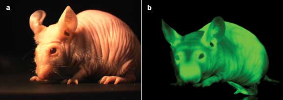 This nude mouse appears normal under bright-field light (a), but it glows under fluorescence excitation with 470-nm blue light (b). Courtesy of AntiCancer Inc.
“Furthermore, the emission spectra of the far-red probes
offers unsurpassed signal-to-noise because these are generally outside of the spectrum
of autofluorescence and do not overlap with conventional blue- or green-emitting
probes,” he added. “New light sources have become available to offer
excitation wavelengths for these far-red probes, and our new optical systems, lenses
and detectors are continuing to allow us to image further into the infrared.”
Published: September 2011