
Fluorescent quantum dots encapsulated in viruses
Method could aid fluorescent tracking
Kevin Robinson
A new era of fluorescent probes for biology was ushered in by GFP. Now researchers
at Indiana University in Bloomington are developing a method for encapsulating
a fluorescent quantum dot inside a virus. The effort could help scientists better
understand the quirky world of viruses, possibly leading to new ways to prevent
viral infection.
Spherical viruses resemble tiny soccer balls.
They’re symmetrical and hollow. The shell of a virus, called the capsid, is
made of protein and encloses the unstructured RNA. Bogdan Dragnea of the university’s
department of chemistry and his colleagues at the University of Massachusetts Amherst
and at Texas A&M University in College Station want to take out the RNA and
put in quantum dots, which are 2 to 4 nm in diameter and grow to 4 to 6 nm once
layers are added to make the surface nonreactive.
“There is plenty of space in
the virus cavity for a quantum dot; however, most of the nucleic acid has to go,”
Dragnea said. Thus, the researchers had to examine which properties are required
for an artificial core to promote the self-assembly of a viral shell around it in
the same way RNA or DNA complexes initiate the self-assembly of a virus in vivo.
For quantum-dot-encapsulated viruses
to work as luminescent markers, they must meet several requirements. Besides promoting
self-assembly of the protein shell, the quantum dots must remain stable and not
release chemically reactive species when exposed to light. The quantum dot and shell
must be water-soluble and stable in a Ph from 5.0 to 7.5.
As decribed in the July 27 online edition
of Nano Letters, the researchers started with brome mosaic virus particles
isolated from infected plants to make the viruslike particle. Dragnea said they
chose that virus because it is well-studied and can be assembled in vitro from proteins
and nucleic acids. “Most importantly, the interaction between the genetic
material and the protein shell is nonspecific,” he said. “This means
that foreign materials are easier to encapsulate.”
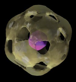
This reconstructed image shows a viruslike particle composed of a
viral protein cage encapsulating a quantum dot. The data was acquired in a transmission
electron microscope at 100 kV and represents the average of ~1000 viruslike
particles.
After dissolving the shell and removing
the RNA using a centrifuge, the researchers introduced various chemically coated
quantum dots to the solution of dissolved shell proteins. They studied several coatings
for quantum dots, including lipid-micelles, streptavidin-biotin-DNA, dihydrolipoic
acid and HS-PEG-COOH.
They assessed the quantum dots’
ability to form a viruslike protein shell and, ultimately, the stability of the
viruslike particles. They used gel electrophoresis to assess the attachment of the
proteins to the quantum dot surface, dynamic light-scattering to determine the hydrodynamic
radii of the quantum dots with various coatings, and transmission electron microscopy
to assess the structure of the self-assembled particles.
To test photostability, they measured
the steady-state luminescence decays of the encapsulated quantum dots. They exposed
the particles to 25 minutes of a Coherent Ti:sapphire laser frequency-doubled to
400 nm and delivering 90 mW at 250 kHz with a pulse length of 200 fs. They split
the beam and used half for a reference beam. Two-pin silicon photodiodes detected
the quantum dot luminescence, and the intensity detected was stored as a function
of irradiation time.
The researchers discovered that the
quantum dots that were coated with HS-PEG-COOH were the most successful. The ones
coated with the lipid-micelles or DHLA did not form capsids at all, and those coated
with streptavidin-biotin-DNA formed capsids, but the capsids broke down, a process
that is made worse by light exposure.
Further examination revealed that the
HS-PEG-COOH-coated quantum dots closely resemble brome mosaic virus particles. They
also were the most photostable, actually gaining luminescence slightly over 13 minutes
of exposure.
“The structure of the viruslike
particle encapsulating a quantum dot is very similar to the structure of native
virus particles isolated from plants,” Dragnea explained. “This means
that the surface coat that we have developed interacts only weakly with the protein
cage, leaving it unperturbed.”
The research may head in several directions,
Dragnea said. The ability of the particles to self-assemble into an icosahedral
(soccer ball) shape opens the possibility of using the technique to grow a variety
of structures, such as three-dimensional crystals. “Meoscopic order at 10
to 100 nm is very difficult to obtain with any other technologies,” he said.
They also will be looking to use the
quantum dot technology for tracking the viruslike particles in cells, where Dragnea
said quantum dots have an edge. “Quantum dots are advantageous with respect
to fluorescent markers because they are more stable and emit stronger.” Lastly,
the researchers likely will continue to tweak the characteristics of PEG-based coatings
for other specific uses.
Published: September 2006