Compiled by Photonics Spectra staff
A new type of genetic tag made by modifying a plant protein has the potential
to illuminate life in never-before-seen detail.
Scientists from the University of California, San Diego (UCSD),
School of Medicine have re-engineered a light-absorbing protein from the cress plant
Arabidopsis thaliana so that when it is exposed to blue light, the altered protein
produces abundant singlet oxygen – a form of molecular oxygen that can be
made visible by electron microscopy (EM).
Development of the small, highly engineered Arabidopsis protein,
dubbed “miniSOG,” may elevate the abilities of EM in the same way green
fluorescent protein (GFP) has made modern light microscopy in biological research
much more powerful and useful, said Nobel laureate Dr. Roger Tsien, Howard Hughes
Medical Institute investigator and UCSD professor of pharmacology, chemistry and
biochemistry.
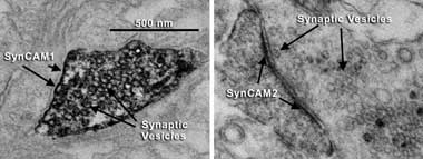
A new electron microscopy technique reveals the previously unknown
locations of two neuronal proteins, SynCAM1 and SynCAM2. The first is an adhesion
protein found at the synapse of neurons sending information. Its relative, SynCAM2,
is used by neurons receiving information. Neurons that send information are distinguishable
because they contain synaptic vesicles, which are used to store neurotransmitters
for communications use. In the images, the vesicles resemble small hollow circles.
Images courtesy of University of California, San Diego, School of Medicine.
“The big advantage of EM is that it has always had much
higher spatial resolution than light microscopy,” Tsien said. “You can
get up to a hundredfold higher useful magnification from EM than from light microscopy.”
He said the result has been extraordinarily detailed 3-D images
of microscopic objects at resolutions measuring in the tens of nanometers –
tiny enough to meticulously render the internal anatomy of individual cells.
Unfortunately, current EM technologies cannot distinguish or highlight
individual proteins in the images. They can be tagged with GFP or other fluorescent
proteins, but they are visible only with the limited resolution of light microscopy.
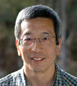
Nobel laureate Roger Tsien.
The new technique, however, enables the scientists to put beacons
on almost any protein to get a snapshot of its location at much higher resolutions
of EM, Tsien said. To demonstrate this ability, the team began working with a protein
from Arabidopsis, a small flowering plant that has long been used as a research
model. Absorbing incoming blue light, the original protein triggers biochemical
signals that inform the plant how much sunlight it receives. The team engineered
the protein so that it could change incoming blue light into a little bit of green
fluorescence and singlet oxygen.
The scientists then used established methods to convert the singlet
oxygen production into a tissue stain that is visible under the EM. They tested
the modified protein’s utility as an EM marker by first using it to confirm
the locations of several well-understood proteins in mammalian cells, nematodes
and rodents. Secondly, they used miniSOG to successfully tag previously unknown
locations of two neuronal proteins in mice.
The scientists are optimistic that miniSOG will enable researchers
who investigate cellular and subcellular structures to find answers to questions
that previously were impossible to ask. The work was reported in the April 5, 2011,
issue of Public Library of Science Biology (doi: 10.1371/jounal. pbio.1001041).