HANK HOGAN, CONTRIBUTING EDITOR
Finding a solitary cancer cell in the midst of millions of healthy ones is no small feat. But that’s what researcher Andrew Filby does, thanks to a camera system that is capable of streaming at nearly 1000 fps and is paired with multiple frame grabbers.
Filby is the faculty lead for the Innovation, Methodology and Applications research theme at Newcastle University in England. “I lead a team of technical specialists who develop and apply different single-cell analysis technologies and methodologies to help answer fundamental questions about human health, disease, and development,” he said. “For several years, we have been interested in approaches that can image cells as they flow past a sensing point, or imaging flow cytometry, at very high speeds.”
The goal is to be able to look at the cells as they stream by and classify them in various ways, such as benign, malignant, healthy, and diseased. This information could then form the basis for an assessment of a patient’s health or a diagnosis for cancer or another disease.
Spotting diseased cells, which can be few in number, in a swarm of healthy cells flowing past is like finding a black swan. “If we think of circulating tumor cells, they can be as low [in number] as one cell in tens of millions,” Filby said.
One current method of imaging cells involves labeling them with special antibodies that have been tagged with fluorochromes. When illuminated with the right light, the fluorochromes glow, allowing the cells to be seen. The antibodies attach themselves to specific classes of cells, such as those that have a cancer marker.
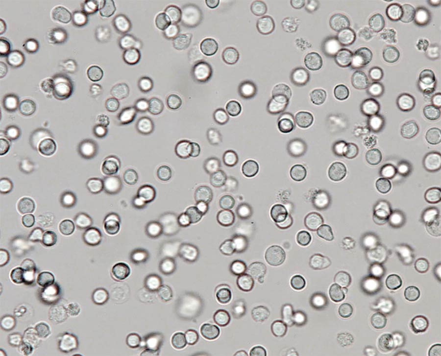
A high-speed camera and frame grabbers produce clear pictures that can be used to detect cancer or other diseases in blood cells. Courtesy of Vision Research Ametek
This approach is expensive and slow, involving multiple steps and a system equipped with lasers of several different wavelengths. Also, researchers and clinicians must have an idea of what they’re looking for, because the tagged antibodies are specific to a given marker or subset of cells.
Filby is developing a method to achieve the same capability as the above approach, but with label-free
imaging. To reach the necessary throughput, he needed a camera and interface with the right characteristics
in terms of speed, sensitivity, and resolution.
950 fps
Enter a proof-of-principle setup consisting of the Phantom S990 camera from AMETEK’s Wayne, N.J.-based Vision Research division, and Coaxlink Octo frame grabbers from Euresys of Angleur, Belgium. The camera streams 9-MP images at almost 950 fps, producing about 9 GP/s.
Getting this much data off the camera quickly enough requires as many
as four CoaXPress CXP-6 frame grabbers. Software from Euresys stitches all of the data from the separate frame grabbers together so that, to the end user, it appears to be a single image.
‘As high-speed camera technology improves and can be
miniaturized, we hope to see label-free, image-based
point-of-care devices developed that can provide robust
results, particularly in resource-poor settings.’
— Andrew Filby, faculty lead for the Innovation, Methodology
and Applications research theme at Newcastle University
The camera is part of a new product line from AMETEK’s Vision Research business unit, according to Kyle Gilroy, a field applications engineer at the company. High-speed cameras can store data locally, which is transferred later for analysis. Alternatively, as is the case with the Phantom series, high-speed cameras can stream the data off so that end users can do what they want with the data, including conducting long recording times or real-time analysis.
Flow cytometry imaging imposes a few requirements, Gilroy said. “You want a lot of sharp images very fast. And you need really high frames rates and short exposure times.”
Together, the camera and frame grabbers satisfy these and other needs. Filby has been working with the technology and refining the solution. The final system may include neural networks that will classify the cell images, perhaps using identifiers developed during the last seven years by the researchers, who are correlating classification from the flow cytometry approach along with the cell categorization from the fluorescent antibody approach.
The initial data from the high-speed flow cytometry imaging is promising, Filby said, with researchers reporting that they like the simplicity of the
system and the image quality, considering the high speed of the cells flowing by.
This proof of the vision-based concept in a research setting is only the beginning, Filby said. “As high-speed camera technology improves and can be miniaturized, we hope to see label-free, image-based point-of-care devices developed that can provide robust results, particularly in resource-poor settings.”