ZURICH, Dec. 31, 2025 — A hybrid system uniting the imaging capabilities of confocal microscopy with the sensitivity of atomic force microscopy has provided new insights into the way influenza enters cells during infection. For this process is not a one-sided occurrence with an invader overwhelming its victim; but rather, images captured in a Petri dish reflect an active interaction between the virus and its host.
A team of researchers from ETH Zurich and Hokkaido University used their setup to track Influenza A virus particles as they traveled along the surface of cells, searching for an adequate collection of receptors. They specifically focused on the interactions of specific proteins including the viral protein hemagglutinin, which binds to the sialic acid on the cell surface, and is then cleaved by another viral protein called neuraminidase, which allows the process to replicate itself elsewhere. Ultimately, this activity precipitates endocytosis, the process by which nutrients (or viruses) are absorbed into a cell.
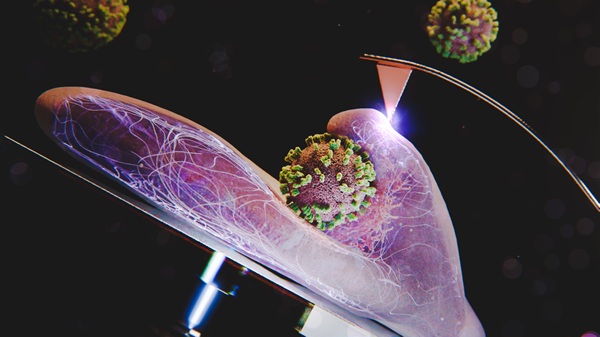
An illustration of a cell absorbing an influenza particle. Courtesy of Emma Hyde/Yohei Yamauchi.
Historically, technologies used independently to study influenza infection, including atomic force microscopy, electron microscopy, and fluorescence microscopy, failed to achieve the level of resolution (particularly spatial resolution) needed to isolate such details.
To adequately isolate these interactions, the instrumentation used by the aforementioned team included a thin, soft-tipped cantilever probe (struck by a laser) to map the cell surface in the performance of atomic force microscopy. Simultaneously, a confocal laser scanning microscope was put to use, which consisted of an electric inverted fluorescence microscope, two lasers (473 and 559 nm) and two fluorescence channels. The objective lens was mounted on an XY movable stage to move in conjunction with the cantilever as the virus was introduced to Madine-Darby canine kidney cells, along with a variety of other cell lines, with essentially the same results observed in each.
“Much of the process of absorption of influenza virus was known, but our perception was that the virus was essentially a bowling ball that ultimately sank into the cell it was infecting,” said Yohei Yamauchi, a professor of molecular medicine at ETH Zurich. “But once we examined the cell surface at nanoscale resolution, we found it was a little more complicated than that. The cell essentially reached out to envelop the virus, which leads to the analogy of the Trojan horse, where the cell thinks it’s getting something good, that it’s not.”
The hybrid system captured images every five seconds, which were then analyzed with data analysis software. The next step in the research, said Yamauchi, will be building a speedier imaging system, and the examination of how different types of influenza virus are absorbed into cells. While the Influenza A viral particles are round, other types are filamentous in shape. And in the future, the hybrid system could be used to examine the effects of drug therapies at the level of cellular interaction.
“With this technology, we get a bird’s-eye view of the cell surface, and how antiviral drugs affect biology,” Yamauchi said. “The cell surface is a stormy sea of activity, but when an inhibitory drug is used, it looks like a very serene sea. It gives you a whole different perspective on what could be going on there.”
The research was published in PNAS (www.doi.org/10.1073/pnas.2500660122).