
Hybrid Imager Spots Elusive Cancer
A combination of three previously unrelated imaging tools promises to lead the way to diagnoses of early-stage ovarian cancer in high-risk women through minimally invasive surgery. The new technique may be better than the current standard procedure of preemptively removing the ovaries.
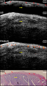
Co-registered images of malignant ovarian tissue obtained with the hybrid imaging device. From top to bottom: OCT image, ultrasound image, superimposed photoacoustic and ultrasound image, and corresponding histology. Yellow diamond arrow = malignant tissue. (Image: University of Connecticut/Biomedical Optics Express.)
Ovarian cancer has a low survival rate because a lack of reliable screening techniques usually means that the disease remains hidden until the later stages. Using multiple imaging tools can help identify earlier the tissue irregularities that signal cancer.
For their diagnostic device, researchers from the University of Connecticut and from the University of Southern California in Los Angeles combined the contrast provided by photoacoustic imaging, the high-resolution subsurface imaging provided by optical coherence tomography (OCT) and the deeper tissue imaging provided by pulse-echo ultrasound.
They tested their device, described in the September issue of Biomedical Optics Express, by imaging both pig and human ovarian tissue. The tripartite technique correctly identified malignant tumors that were later confirmed by staining the tissue and examining it under a microscope. These initial tests were performed on tissue that had been surgically removed, but the diameter of the device — 5 mm — is small enough that it could be inserted through a small slit to image tissue in live patients.
For more information, visit: www.uconn.edu
Published: September 2011