Single plane illumination microscopy is an imaging technique widely used in biomedical research, allowing for high-speed, deep imaging that provides volumetric data.
ELISABETH KUGLER, LUXENDO GMBH AND ZEEKS - ART FOR GEEKS LTD, MALTE WACHSMUTH, LUXENDO GMBH, AND DIMITRI KROMM, DELFT UNIVERSITY OF TECHNOLOGY
Light-sheet fluorescence microscopy, also called single plane illumination microscopy (SPIM) is a noninvasive 3D imaging method that employs a sheet of light to illuminate a thin plane of a specimen. This technique reduces out-of-focus light and allows deep tissue imaging with reduced photodamage. This means that SPIM is particularly well suited for in vivo observation at subcellular resolutions that yield only minimal phototoxicity and fluorophore bleaching. Due to these properties, SPIM has become widely used for dynamic studies with time-lapse imaging of live biological samples over extended periods of time (hours to days).
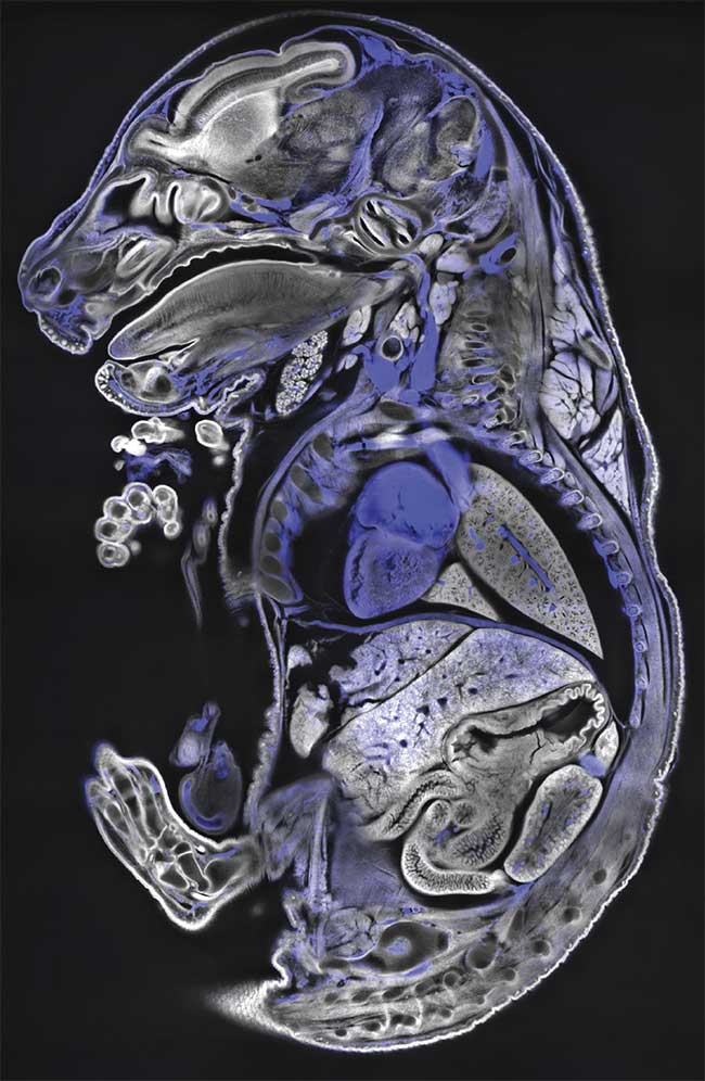
Methylene blue imaging in a mouse sample with the Bruker Luxendo LCS SPIM. Courtesy of Montserrat Coll Llado/European Molecular Biology Laboratory.
Together, the ability to capture high-resolution 3D data deep within tissues while preserving sample integrity has made it an indispensable tool for modern biomedical research. For example, SPIM is used to study intracellular dynamics in single cells in vivo or follow single cells and tissues in developing embryos of flies, plants, fish, mice, chickens, and many other organisms. Additional application areas include the functional imaging of brains and hearts, where rapid volumetric recordings are required. A more recent use of SPIM is the anatomical/histological imaging of larger, centimeter-sized organisms and tissues, such as mouse brains or human cancer tissues — a realm that largely profits from increased acquisition speeds.
There are numerous advantages to SPIM imaging for the researcher, but it is important to explore geometries of the instrumentation, address data challenges, and consider current and future innovation and how that may affect the long-term use of SPIM.
Shedding light on samples
SPIM imaging offers several advantages beyond reduced phototoxicity and its inherent outstanding time-lapse capacities. Some of these advantages include:
1) Imaging speed: Rapid imaging allows scientists to capture dynamic processes with sufficient time sampling as well as to acquire data from large or multiple samples in short time frames. Examples include calcium imaging in the brain or path-seeking in macrophages.
2) Large field of view (FOV) and depth penetration: SPIM enables the capturing of detail in entire organs and organisms. The ability to study detail in their 3D environment with respect to the whole organ/organism context allows for new insights and discovery, such as the discovery of blood vessel cell behaviors1. In the emerging field of cleared sample imaging at use in neuroscience and developmental biology, specimens in the centimeter range have been successfully imaged.
3) Sample chambers: SPIM’s sample chamber creates a microenvironment with tight sample control, often multiple illumination and view angles, and stable physiological conditions. It accommodates interventions and sample manipulations that might be carried out during observation, such as anesthesia, drug responses, and hypoxia studies.
4) Versatile sample mounting: SPIM allows versatile sample mounting, catering to various specimens and experimental setups. Examples range from zebrafish larvae embedded in gel with capillaries, uterus-like microenvironments, to cleared mouse brains covered with tissue-clearing solutions inside of specialized cuvettes. These mounting methods might include gel embedding with support from above/below, flat mounting, hooking, and (microfluidic) flow channels.
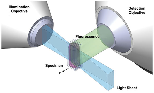
This rendering shows the orthogonal illumination of a cylindrical sample (magenta) with a sheet of light originating from the illumination objective lens (left). The emitted light is detected by a detection objective lens (right). In classical selective plane illumination microscopy (SPIM) setups the sample can be translated in 3D, as well as rotated. Courtesy of Dimitri Kromm.
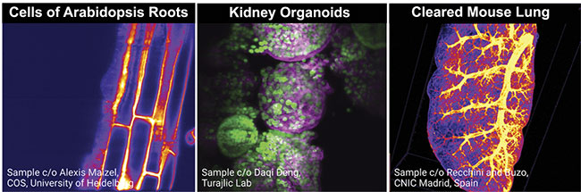
This image shows a selection of different samples imaged with selective plane illumination microscopy (SPIM), including cells of plant roots, kidney organoids, and cleared mouse lungs. Courtesy of Elisabeth Kugler.
SPIM geometries and platforms
Prior to using SPIM for a project, in a laboratory, or in a facility, one should carefully assess the requirements for the intended applications. If SPIM is chosen, the microscope design concept influences all work associated with the experiment, whether open source, commercial, or custom built. Factors to consider include sample mounting, system alignment/maintenance, data formats, sizes, processing steps, etc.
Characteristics of the sample
The best microscope is of little use if a consistent optimized mounting method is not established. Problems arise from various components, such as sample drift, refractive index mismatch, or sample death. Proper mounting, however, depends on the nature of the sample itself, along with the configuration of the microscope, environmental factors, and optical requirements of the system. For example, mammalian samples require sterile controlled sample chambers at ~37 °C and their comparably slow development calls for time-multiplexed imaging of several samples. Intracellular dynamics, on the other hand, require high spatiotemporal resolution, for which gels for embedding can cause unwanted optical aberrations.
Customized or commercial
It might be tempting for many technology-savvy scientists to build their own microscope. However, there are potential challenges in doing so, and a plethora of systems have been developed over 20 years.
When choosing a SPIM platform, be it commercial, self-built, or open-source, several factors must be considered, such as budget, time, ease of use, flexibility, and longevity.
Typically, commercial systems are the shortest route to allow multiple users to generate reproducible high-quality data, assuming funding is available for the purchase. If, however, unique requirements exist for resolution, FOV, sample mounting (flexibility), etc., an open-source or self-made design might be the right choice. Building a system on your own will require advanced optics knowledge, optics-related tools, and ideally a well accessible workshop for custom parts and prototyping.
Open-source solutions such as Open-SPIM3 or the Meso-SPIM4 initiative support inexperienced newcomers and minimize the number of custom parts needed. For example, files for 3D printing are openly shared and combined with best-practice forum discussions. Another new approach is provided by the Flamingo project5 from the Huisken group, which allows users to apply with a project and receive a light-sheet microscope for temporary usage. This means that the SPIM comes to the scientists’ lab, and experiments can be performed with a temporarily available microscope.
SPIM illumination and detection of light generally happens in decoupled optical paths and creates space for a variety of geometrical arrangements and objective lens combinations. Typically, the nature of the sample dictates the microscope geometry to be used. The sample size, number of specimens, access to the sample, or even the media can affect the choice of geometry.
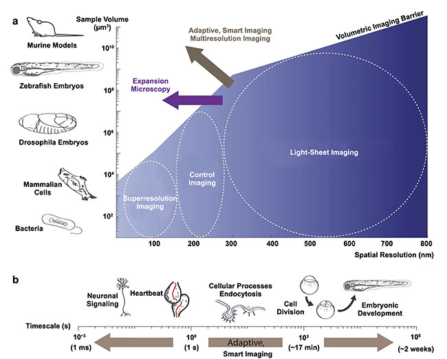
An illustration displaying how a variety of imaging technologies can be put to work surrounding common biological samples. The illustration also sign-posts to the current developments and future leading edges of selective plane illumination microscopy (SPIM) developments, including expansion microscopy and adaptive, smart, multiresolution imaging. Adapted with permission from Reference 2/CC BY 4.0.
Data processing challenges
As imaging methods evolve, more data is generated and new data types evolve, necessitating scalable data handling solutions. Particularly with long-term timelapses and multimodal data, scientists face new challenges with data processing, sharing, and storage (on both the hardware and software sides).
The development of open-source software packages tailored for SPIM processing has democratized access to advanced processing tools. Examples of processing methods that have been driven by the open-source community include 3D stitching, registration, fusion, and deconvolution, interactive object tracking during acquisition, and virtual 3D reconstructions.
While the SPIM community is very active in open-source software development, there are powerful commercial software solutions available that provide the most-needed functionalities required for SPIM data processing that are listed above. Examples available on the market are Imaris (Bitplane), Luxendo Image Processor (Luxendo), Arivis Vision 4D (Zeiss), and Huygens (SVI).
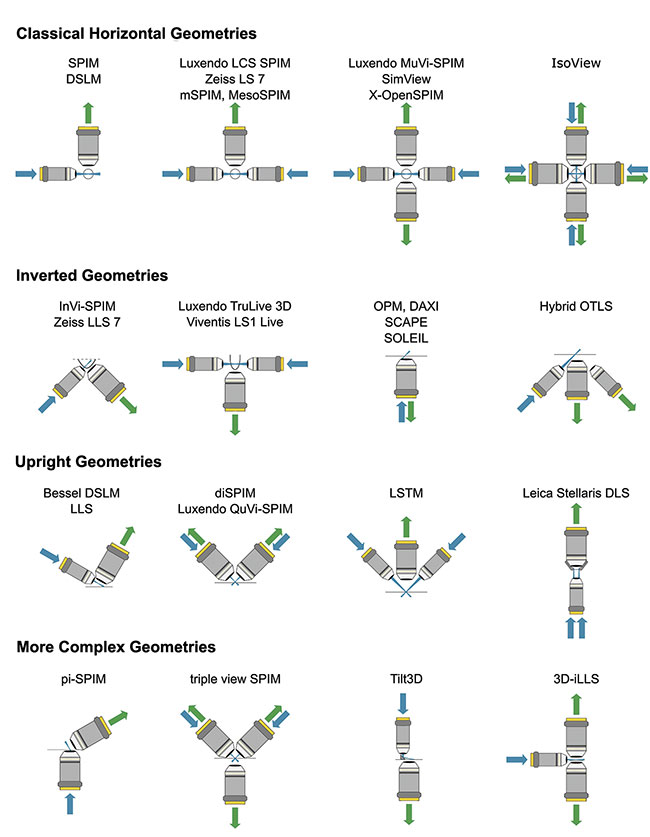
An overview of commonly used and more sophisticated selective plane illumination microscopy (SPIM) geometries of home-built, open-source, and commercial light-sheet microscopes. DLS: dynamic light scattering; DSLM: digital scanned laser light-sheet fluorescence microscopy; OTLS: open-top light-sheet. Adapted with permission from Reference 6. Courtesy of Dimitri Kromm.
With the evolution of data, the datatypes in biomedical image analysis also change. In addition to proprietary data formats, open-source, partially streaming-enabled data types have emerged or are being developed that allow the user to store multidimensional, complex pixel and voxel values, as well as metadata.
Given the data volume, efficient data compression is crucial for managing storage and facilitating data transfer. Some of the most common approaches include lossless compression, lossy compression, predictive coding, sparse representation, region-of-interest compression, lossy to lossless progressive compression, cropping, preprocessing, down-sampling, and domain-specific compression.
Together, with new and evolving data, data challenges will require hardware and software to develop with it.
Future of SPIM
SPIM is an ever-evolving field and many aspects, such as sample, geometry, medium, and illumination beam shapes, are still evolving. There are some exciting frontiers that have just begun to be explored.
Different illumination beam variations of SPIM have emerged, each optimized for specific applications. For example, in recent years, an evolution from Gaussian via “self-healing” Bessel to lattice light sheets has occurred. The user is given the opportunity to increase axial resolution with the latter two light-sheet variants and their smaller beam-waists at the cost of sacrificing part of the available photon-budget (faster bleaching). Tailored light-sheet shapes can provide new biological insights, i.e., by imaging the previously unresolved.
Spatial resolution is a highly sought-after property, particularly for subcellular studies. Techniques such as lattice light-sheet microscopy and adaptive optics (AO) have been employed to achieve higher resolution imaging of live biological samples. For example, AO is a technique used to mitigate aberrations and optimize image quality in 3D imaging that would be difficult to achieve with traditional instrumentation. By integrating a deformable mirror and wavefront sensing into the SPIM setup, AO dynamically corrects distortions introduced by local and global refractive index variations.
Integrating different imaging modalities, such as fluorescence lifetime imaging (FLIM), imaging fluorescence correlation spectroscopy, or Brillouin microscopy, allows the simultaneous acquisition of different types of information from a sample: For example, combining spatial information derived from SPIM with more functional/dynamic information from FCS, which measures the concentration of molecules based on fluorescent fluctuations. Analysis of these data points can then provide insights into tissue heterogeneity as seen in tumors, which can inform the identification of new biomarkers.
For fixed sample preparation, tissue-clearing methods have enabled imaging of larger samples, such as whole organs and organisms, while the related expansion microscopy achieves resolutions beyond the diffraction limit by expanding the sample isotropically to a multiple of its original size, while maintaining the tissue integrity. In both techniques, chemicals are used to make the samples transparent and homogenize their refractive indices.
Thus, the specimen becomes optically more accessible and when expanded further in size, the resolution effectively improves by the expansion factor. The true elegance of this only becomes apparent when working with larger samples, such as whole organs or organisms, which can be imaged in SPIM, mostly due to the reduced photo-bleaching and high acquisition speeds. For example, combining large-scale SPIM data with high resolution was most recently highlighted by new findings on skull bone marrow immune cells7.
During the past two decades, numerous established model organisms have been imaged live due to the low phototoxicity of SPIM. However, with a wider spread of light-sheet microscopes and experience of the community, it can be expected that entirely new models will emerge that were not previously accessible with other microscopy techniques. This is exemplified by studies of the Erturk lab, which recently showed that tumors are detectable at the single-cell level in whole mice, whereas techniques such as MRI would require large tumor masses to identify such a tumor.
Another example are spheroids and organoids, which are highly sensitive samples that become a more widely used sample type in various fields, such as cancer research, and that benefit from the low phototoxicity of SPIM. Recent studies showed that SPIM can be used to record light-sensitive reef-building corals in 3D, a sample otherwise defective with 3D live imaging.
Such examples show that SPIM is already an important workhorse for imaging in biomedical research and life sciences experiments. Looking to the future, the field will continue to evolve rapidly on many frontiers, be it sample preparation, microscope geometry, illumination concepts, or software.
Meet the authors
Elisabeth Kugler is director at Zeeks - Art for Geeks Ltd and external Marcom specialist for Luxendo GmbH. Her expertise lies in vascular biology, biomedical image analysis, and science communication; email: [email protected].
Dimitri Kromm is coordinator of the Smart Optics Lab at the Delft Center for Systems and Control (DCSC), at Delft University of Technology. He specialized in multiview imaging of live model organisms using light-sheet microscopy and on minimizing the effects of optical aberrations in these systems; email: [email protected].
Malte Wachsmuth is managing director at Luxendo GmbH of Bruker Fluorescence Microscopy, and head of application, support, and service. He is a trained biophysicist with focus on advanced optics and optical applications, particularly light-sheet microscopy; email: [email protected].
References
1. E.C. Kugler et al. (2019). Cerebrovascular endothelial cells form transient Notch-dependent cystic structures in zebrafish. EMBO Rep, Vol. 20, No. 8, p. e47047.
2. S. Daetwyler and R.P. Fiolka. (2023). Light-sheets and smart microscopy, an exciting future is dawning. Commun Biol, Vol. 6,
No. 1, p. 502.
3. P.G. Pitrone et al. (2013). OpenSPIM: an open-access light-sheet microscopy platform. Nat Methods, Vol. 10, No. 7,
pp. 598–599.
4. F.F. Voigt et al. (2019). The mesoSPIM initiative: open-source light-sheet microscopes for imaging cleared tissue. Nat Methods, Vol. 16, No. 11, pp. 1105-1108.
5. Huisken Lab. Flamingo Project, huiskenlab.com/flamingo/.
6. D. Kromm (2023). Pushing Light-Sheet Microscopy to Greater Depths. Doctoral Thesis, Heidelberg University.
7. Z.I. Kolabas et al. (2023). Distinct molecular profiles of skull bone marrow in health and neurological disorders. Cell, Vol. 186,
No. 17, pp. 3706-3725.e29.