Photonics HandbookBioScan
Imaging Techniques Improve Visualization of Vertebrate Specimens
Imaging techniques pioneered by researchers at the University of Kansas (KU) could enable scientists around the world to better capture complex morphological features of vertebrates for comparative anatomical studies.
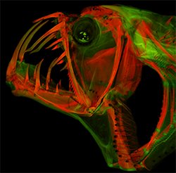
Cleared-and-stained head of a frightening deep-sea predator, Sloan's viperfish (Chauliodus sloani). Photo is under fluorescent light. Courtesy of M. Davis.
The KU team developed two methods for simplifying and improving the digital imaging of vertebrate skeletons and their components. The first method uses fluorescence microscopy to create images of alizarin-stained specimens. Alizarin, a stain used in the clearing and staining process to identify bones in a specimen, fluoresces when exposed to the right wavelengths of light, which can facilitate the identification of skeletal elements in extant and fossil specimens.
“Alizarin red is used to dye a specimen’s bones, and it fluoresces like a Grateful Dead poster,” said professor W. Leo Smith.
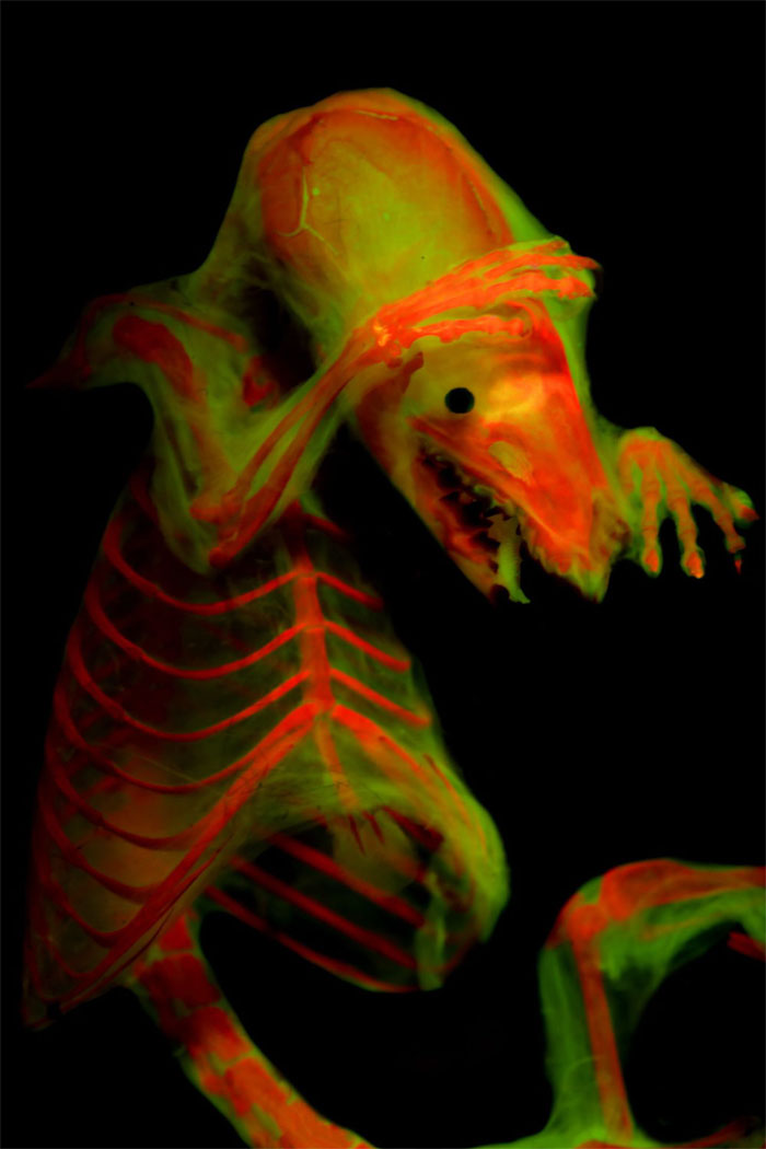
Cleared-and-stained North American least shrew (Cryptotis parva). Photo under fluorescent light. Courtesy of M. Girard.
“We use lights that have high energy and look for reflections of re-emitted fluorescent wavelength, and the microscope has filters that block all the other light,” he said. "The skin and everything else disappears because it doesn’t fluoresce — it’s a fast way to clear out all the extra stuff and is incredibly useful when you're trying to see where bones are connected."
The second imaging method involves the nonpermanent mounting of cleared-and-stained vertebrate specimens in a glycerine-gelatin matrix that allows researchers to temporarily pose specimens for otherwise impossible scientific or artistic images. The team discovered a 40 percent glycerine mixture that held specimens well and was sufficiently translucent for photography, allowing new looks at specimens that could float within the matrix.
“You can see through this medium and give the specimen structure,” Smith said. “Now you can get a photo of a fish specimen head on and look at it from all these different angles.”
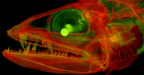
Cleared-and-stained juvenile lancetfish (Alepisaurus) with enormous fangs. Photo is under fluorescent light. Courtesy of M. Davis.
Smith stressed the importance of providing compelling images to convey information to investigators and the public.
“At end of the day, the picture is worth a thousand words,” he said. “Images allow you to fundamentally share how things work and improve your ability to tell someone else about your novel discoveries.”
The research was published in Copeia (doi: 10.1643/CG-18-047).
Published: September 2018