Parents and caretakers of little ones know that babies move around constantly — often getting into trouble in the process. But researchers in and around London know that, as an indicator of overall health, what is going on inside babies’ brains can be just as important as little ones’ physical activity. The researchers used low levels of light and 3D imaging to create pictures of the activity that takes place in the infant brain during development.
Of course, classic imaging technology can be problematic when used for this purpose, given a baby’s propensity for constant movement. Magnetic resonance imaging requires a child to remain still, and near-infrared spectroscopy, which has been used to test children’s responses to cues such as sounds or colors, has low spatial resolution. Parents know that babies are usually busy exploring — out of curiosity rather than science. The challenge, therefore, was to develop a mechanism to accommodate movement, while at the same time getting precise measurements of blood flow and other brain activity — in short, designing equipment that could take measurements through every crawl, roll, and squirm.
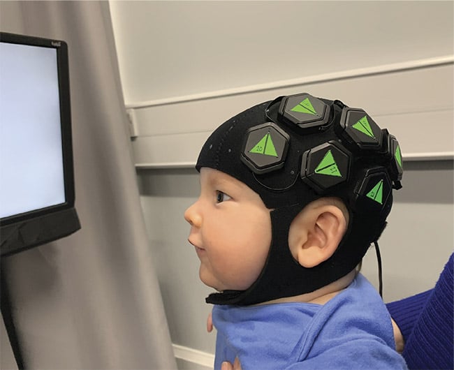
A baby wears a cap designed by scientists at Cambridge University to provide images of brain activity during
development. Courtesy of Robert Cooper.
A team that drew scientists from a variety of institutions — including University College London (UCL); the Rosie Hospital of Cambridge University; and Gowerlabs Ltd., a spinoff company that worked on the technology — created a wearable cap that fits on a baby’s head and uses high-density diffuse optical tomography to monitor activity in the brains of 6-month-old infants. The technique is an optical imaging modality that uses near-infrared light to measure tissue oxygenation, which marks where activity is occurring in the brain.
“Diffuse optical tomography is inherently very safe,” said Robert Cooper, an EPSRC (Engineering and Physical Sciences Research Council) Early Career Fellow at UCL, who led the project. “Attaching fibers to infants’ heads is hugely difficult and constraining. This new generation of technologies allows us to provide high-density sampling without the burden of fibers.”
The flexible neoprene (synthetic rubber) cap was tested on 20 healthy infants. The cap uses LEDs that deliver red and near-
infrared light, and optical detectors that are laid out in sensor tiles arranged over the child’s scalp. A series of docks are distributed throughout and connected to a PC that provides power.
“The system Gowerlabs has built has a beautiful system architecture,” said Cooper. “It is fully modular and everything is in place to scale the system up to cover the entire scalp, which will need somewhere between 24 and 56 modules, depending on
the size of the head. Right now the most we could apply was
12 modules, so we shared them equally over two hemispheres.”
Researchers showed the infants a series of prompts that each lasted several seconds, including videos of actors mouthing words and nursery rhymes, or making hand gestures, such as the ever-popular peekaboo. The majority of the babies in the experiment reacted for a sufficient amount of time for the cap to provide scientists with measurements of oxyhemoglobin and deoxyhemoglobin responses at defined points in the array.
In time, the researchers hope that the technology will help to capture images of developmental issues associated with autism spectrum disorders and cerebral palsy. But for now, the fact this technology can accommodate babies’ tendencies to move around during an examination is a breakthrough — for scientists and for parents.