While imaging cells using real-time fluorescence microscopy methods, professor Kaoru Tamada of Kyushu University’s Institute for Materials Chemistry and Engineering and her group found that they could improve resolution under a conventional widefield microscope close to the diffraction limit by simply changing the surface beneath the cells.
The researchers demonstrated that using a glass surface embedded with self-assembled gold nanoparticles can improve resolution of conventional microscopes and image living cells at high speeds.
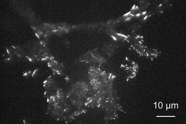
A mouse fibroblast cell imaged on a metasurface made of gold nanoparticles under a total internal reflection fluorescence (TIRF) microscope exhibits enhanced and confined emission from light-emitting paxillin protein at focal adhesions near the membrane. Courtesy of Kaoru Tamada, Kyushu University.
The method allows for imaging close to the refraction limit without high-powered lasers, which can destroy living cells, and/or the scanning of the sample or processing of multiple images, which can inhibit real-time imaging.
“Recent techniques can produce stunning images, but many of them require highly specialized equipment and are incapable of observing the movement of living cells,” Tamada said.
Fluorescence microscopy involves tagging cell structures with molecules that absorb energy from incoming light, and, through the process of fluorescence, re-emit the energy as light of a different color; that light then forms an image.
While cells in the method are typically imaged on plain glass, Tamada’s group coated the glass surface with a self-assembled layer of gold nanoparticles, itself covered with a thin layer of silicon dioxide. This created a metasurface with the optical properties necessary to allow it to collect energy from nearby light-emitting molecules for highly efficient re-emission, thereby producing enhanced emission confined to the 10-nm-thick nanoparticle surface. The organized metal nanoparticles, meanwhile, exhibited localized surface plasmon resonance, which allows the metasurface to achieve the required functionality.
“By introducing the nanoparticles, we have effectively created a light-emitting plane that is only several nanometers thick,” Tamada said. “Because the light of interest is emitted from such a thin layer, we can better focus on it.”
According to Abbe’s diffraction limit, the metasurface’s high refractive index improves resolution, and the quick energy transfer to the metasurface allows for further localized emission points by reducing diffusion.
Using the metasurface, the researchers were able to image, in real time, mouse cells known as 3T3 fibroblasts engineered to produce paxillin. A protein, paxillin is modified to emit green light when excited.
By illuminating the whole sample with laser light perpendicular to the surface, the researchers were able to image changes in paxillin near the cell membrane.
Tilting the light source to achieve total internal reflection, the researchers were additionally able to achieve even higher contrast images. Here, most of the light was reflected off the surface, with only a small amount reaching the cell side, reducing stray emission produced by illumination penetration deep into the cell.
Analyses of images recorded every 500 milliseconds with a superresolution digital camera showed clear differences in intensity over spots covering only a few pixels, indicating that the resolution was approximately 200 nm, which is close to the diffraction limit.
In an instance in which less input energy is necessary, thereby reducing cell damage over time, the researchers were also able to image cells for longer on the metasurface.
“Metasurfaces are a promising option for improving resolution for researchers around the world using conventional optical microscopes that they already have,” Tamada said.
The research was published in ACS Applied Nano Materials (www.doi.org/10.1021/acsanm.0c02300).