
Intelligent Microscope Framework Captures Biological Events
LAUSANNE, Switzerland, Sept. 13, 2022 — Biophysicists at École Polytechnique Fédérale de Lausanne (EPFL) introduced a method to automate microscope control for imaging biological events in detail with the help of artificial neural networks, while limiting stress on the sample. The team applied its development to the technique of fluorescence microscopy.
Fluorescence microscopy can be used to collect data on specific biological events, though the event-specific content that can be collected from a sample is limited. The process can be time-consuming — it can take hours for one bacterium to divide — and indiscriminate imaging poses other challenges. Indiscriminate image capture and capturing images as frequently as possible use excessive light, which can deplete the fluorescence from a sample faster and can prematurely destroy living samples. Photobleaching and phototoxicity constrain imaging speed and duration.
Further, these approaches are apt to generate uninteresting images, since only a few would contain images of dividing bacteria.
Using the method, the team captured mitochondrial and bacterial divisions at imaging rates that match their dynamic timescales while extending overall imaging durations.
“Because event-driven acquisition allows the microscope to respond specifically to complex biological events, it acquires data enriched in relevant content,” the researchers said.
The EPFL method adapts acquisitions on the fly by switching between a slow imaging rate while detecting the onset of events, and a fast imaging rate during their progression.
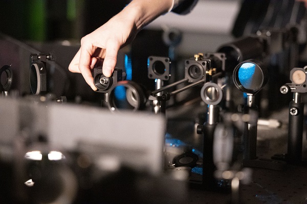
EPFL researcher Suliana Manley’s fluorescent microscope in the Laboratory of Experimental Biophysics. A method to automate microscope control for imaging biological events in detail adapts acquisitions on the fly by switching between a slow imaging rate while detecting the onset of events, and a fast-imaging rate during their progression. The method overcomes limitations to conventional manual image capture. Courtesy of Hillary Sanctuary/EPFL.
“An intelligent microscope is kind of like a self-driving car,” said investigator Suliana Manley of EPFL’s Laboratory of Experimental Biophysics. “It needs to process certain types of information, subtle patterns that it then responds to by changing its behavior. By using a neural network, we can detect much more subtle events and use them to drive changes in acquisition speed.”
Manley and her colleagues first solved how to detect mitochondrial division, which is more difficult than for bacteria such as C. crescentus. Mitochondrial division is unpredictable because it occurs infrequently and can happen almost anywhere within the mitochondrial network at any moment.
The scientists trained the neural network to look out for mitochondrial constrictions, a change in shape of mitochondria that leads to division, combined with observations of a protein known to be enriched at sites of division. When both constrictions and protein levels are high, the microscope switched into high-speed imaging to capture many images of division events in detail. When constriction and protein levels are low, the microscope switched to low-speed imaging to avoid exposing the sample to excessive light.
With the intelligent fluorescent microscope, the scientists showed that they could observe the sample for longer compared to standard fast imaging. While the sample was more stressed compared to standard slow imaging, the team obtained more meaningful data.
“The potential of intelligent microscopy includes measuring what standard acquisitions would miss,” Manley said. “We capture more events, measure smaller constrictions, and can follow each division in greater detail.”
The scientists are making the control framework available as an open source plug-in for the open microscope software Micro-Manager, with the aim of allowing other scientists to integrate artificial intelligence into their own microscopes.
The research was published in Nature Methods (www.doi.org/10.1038/s41592-022-01589-x).
Published: September 2022