
Intravital Imaging Provides Insight into Liver Disease Progression
The high-resolution, 3D-viewing capabilities provided by a new microscopy method from the Korea Advanced Institute of Science and Technology (KAIST) could lead to insights into nonalcoholic fatty liver disease (NAFLD), a condition in which too much fat is stored in the liver. The researchers used their technique to observe how tiny droplets of fat, or lipids, accumulated in the liver cells of living mice over time.
“It has been challenging to find a treatment strategy for NAFLD because most studies examine excised liver tissue that represents just one time-point in disease progression," professor Pilhan Kim said. “Our technique can capture details of lipid accumulation over time, providing a highly useful research tool for identifying the multiple parameters that likely contribute to the disease and could be targeted with treatment."
To capture the dynamics of NAFLD in living mouse models, the researchers developed a custom intravital, confocal, two-photon microscopy system that acquires images of multiple fluorescent labels at video-rate and with cellular resolution.
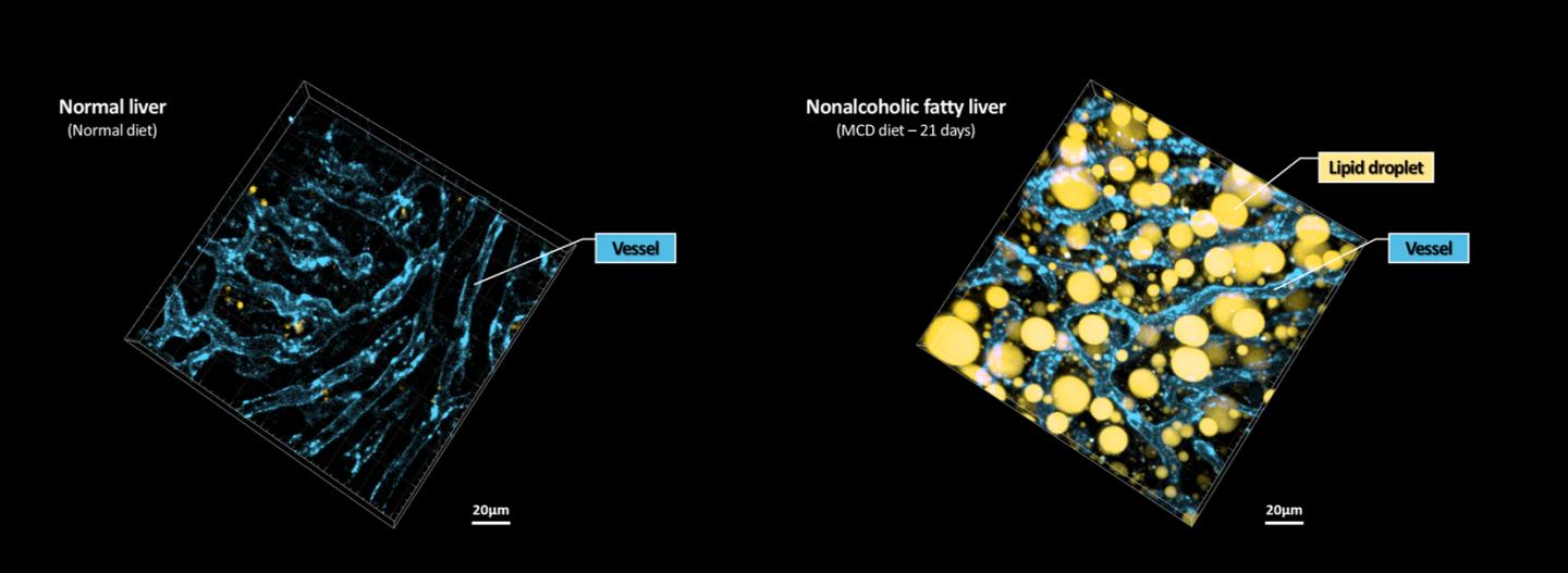
Researchers developed a way to visualize the progression of nonalcoholic fatty liver disease (NAFLD) in mouse models of the disease. Using the technique, they captured lipid droplet accumulation (yellow) and microvasculature (blue) in the livers of normal mice and mice with NAFLD induced with a methionine- and choline-deficient diet. Courtesy of Pilhan Kim, Korea Advanced Institute of Science and Technology.
A polygonal mirror that rotates at more than 240 mph is used to provide extremely fast laser scanning for acquiring images quickly. Four different lasers and four high-sensitivity optical detectors allow the acquisition of multicolor images so researchers can identify different-colored fluorescent probes used to label the lipid droplets and microvasculature in the mice livers.
With video-rate imaging capability, the continuous movement of liver tissue in live mice due to breathing and heart beating can be tracked in real time and precisely compensated, Kim said. "This provided motion-artifact-free, high-resolution images of cellular- and subcellular-sized individual lipid droplets,” he said.
The researchers used their approach to observe the development and spatial distribution of lipid droplets in individual mice with NAFLD induced by a methionine- and choline-deficient diet. Using the nonalcoholic steatosis and steatohepatitis mouse model, the researchers longitudinally visualized and quantitatively analyzed the development of lipid droplets at a subcellular resolution during the progression of NAFLD up to 21 days in vivo.
The researchers will use their technique to visualize various immune cells and lipid droplets to better understand the complex liver microenvironment in NAFLD progression.
“Our approach can capture real-time changes in cell behavior and morphology, vascular structure and function, and the spatiotemporal localization of biological components while directly visualizing lipid droplet development in NAFLD progression,” Kim said. “It also allows the analysis of the highly complex behaviors of various immune cells as NAFLD progresses.”
The research was published in Biomedical Optics Express (www.doi.org/10.1364/BOE.3958).
Published: September 2020