An imaging system called cryo-micro-optical sectioning tomography (cryo-MOST) could help speed new drug development for Alzheimer’s disease by offering a better way to monitor the brain changes indicative of Alzheimer's in mouse models. Cryo-MOST provides micron-level, 3D visualization of protein deposits, known as senile plaques, that are characteristic of the disease in humans and mice.
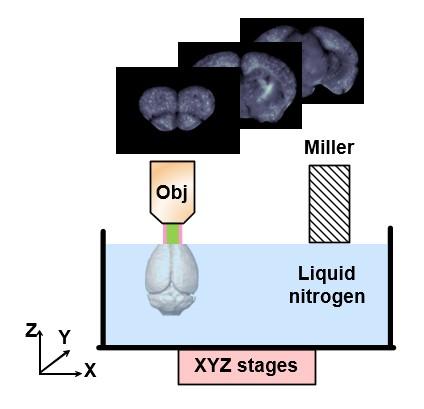
The cryo-MOST imaging system allows whole-brain mapping of senile plaques in the mouse brain. The system cools the sample by keeping it immersed in liquid nitrogen to enhance fluorescence from senile plaques. For whole-brain imaging, the XYZ stages move the sample to automatically alternate between imaging and the miller machine that removes a layer of the tissue each time the surface is imaged. Courtesy of Jing Yuan, Huazhong University of Science and Technology.
Using cryo-MOST, researchers from Huazhong University of Science and Technology were able to obtain whole-brain coronal distribution of senile plaques in a transgenic mouse without exogenous dye. A label-free imaging system, cryo-MOST takes advantage of the natural fluorescence of senile plaques after exposure to excitation light.
Researchers found that by lowering the tissue temperature to less than −100 degrees Celsius, they could brighten the autofluorescence from the plaques and improve the resulting images. Validation using immunofluorescence demonstrated the capacity of cryo-MOST to visualize and distinguish senile plaques with a high degree of sensitivity and spatial resolution.
“Exogenous dyes may lead to unspecific, false-positive or uneven labeling in brain tissue, which impedes observation of authentic pathological structure changes of Alzheimer’s disease,” said researcher Jing Yuan. “Our label-free approach avoids these problems while also simplifying the sample preparation, thus accelerating the research process.”
According to researchers, compared with imaging in room temperature, cryo-MOST provides better signal intensity and signal-to-noise ratio.
To maintain the ultralow temperatures required to enhance the fluorescence, the team created a system that allowed imaging of a sample immersed in liquid nitrogen. Because traditional optical microscopy can only image the surface of the tissue, they incorporated a mechanical milling machine that removed a layer of the tissue each time the surface was imaged. When acquiring images of the whole mouse brain, the system automatically alternated between milling and imaging by using a mechanical stage to move the sample.
To demonstrate the ability of cryo-MOST to image the brain-wide distribution of senile plaques, the researchers used it to image a whole brain from a 17-month APP/PS1 mouse model of Alzheimer's disease. A lateral resolution of 1.072 microns and an axial resolution of 17.152 microns were used to detect fluorescence at a wavelength of 536 nm. The researchers say that these parameters could be improved by using a better microscope.

“The images from an aged Alzheimer’s disease mouse revealed that senile plaques have spread to the whole brain,” said Yuan. “This indicates that the disease not only hurts memory and intelligence, but may also cause an overall deterioration of other brain functions.”
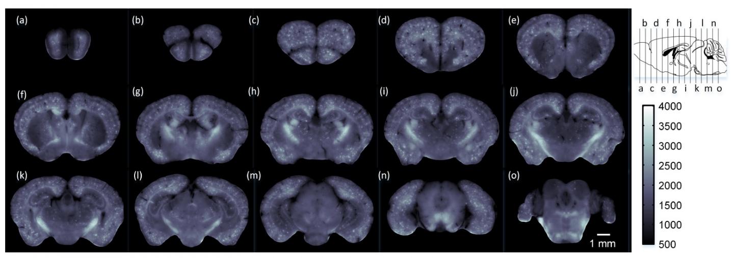
Using cryo-MOST, the researchers acquired a brain-wide map of an Alzheimer mouse model showing that senile plaques had spread to the entire brain. The images are at 1-millimeter intervals from the olfactory bulb to the cerebellum. The specific locations are shown in the upper right insert. Courtesy of Jing Yuan, Huazhong University of Science and Technology.
Because the size of the tissue imaged is limited only by the maximum moving range of the mechanical stage, the system could be used for studying human brain tissue from deceased donors. It could also be useful for visualizing other biological molecules that exhibit autofluorescence in other organs. For example, it could image metabolism distribution in organs such as the kidney and liver.
The research team is currently working to improve the automation of the approach to speed up image acquisition and is incorporating enhancements that will improve image quality. These upgrades could allow their approach to be used for more applications. The researchers are also continuing to use cryo-MOST to study senile plaque distribution and morphology changes that come with the progression of Alzheimer’s disease.
According to researchers the cryo-MOST system is simple and efficient compared with conventional approaches and because it is optical, offers more detailed information than other imaging techniques such as magnetic resonance imaging (MRI) or Positron Emission Tomography (PET).
Label-free brainwide visualization of Alzheimer’s pathology, such as cryo-MOST provides, could potentially be useful for understanding neurodegenerative disease mechanisms and evaluating drug efficacy.
Yuan said, “In addition to evaluating drug efficacy for neurodegenerative diseases, we believe our method will help neuroscientists understand the relationship between senile plaque distribution and other key factors of Alzheimer's disease. This will enhance the comprehensive understanding of the disease mechanisms and treatment.
“Studying the brain-wide distribution of senile plaques in mice will facilitate an understanding of how brain functions deteriorate during Alzheimer's disease progression,” he said. “We hope that cryo-MOST will accelerate the development and evaluation of Alzheimer's disease treatments.”
The research was published in Optics Letters, a publication of OSA, The Optical Society (doi: 10.1364/OL.42.004247).