STORRS, Conn., Feb. 20, 2020 — Guoan Zheng, a University of Connecticut (UConn) professor of biomedical engineering, has published his findings on a successful demonstration of a lensless on-chip microscopy platform in Lab on a Chip. Zheng suggests his platform eliminates several of the most common problems with conventional optical microscopy while providing a low-cost option for the diagnosis of disease.
UConn researchers plan to continue to refine Zheng’s microscopy technology to enhance its use in commercial and clinical applications. Courtesy of Guoan Zheng.
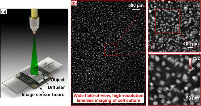
Rather than using lenses to magnify the tissue sample, Zheng’s platform relies on a diffuser that goes between the specimen and the image sensor or camera. The diffuser randomly moves to different positions while the sensor acquires the images, gathering the encoded object information that will later be used to recover an image for viewing by clinicians or researchers.
“Imagine being able to read a whole book at once instead of just a page at a time. That’s essentially what we hope our technology will allow clinicians to do.”
—Guoan Zheng
Zheng implemented an imaging technique known as ptychographic imaging, which typically uses a focused beam to illuminate a sample and then records the pattern created by the diffracted light. To recover an entire complex image, such as a tissue sample, ptychography requires thousands of patterns to be recorded while scanning the sample to different positions.
“Although ptychography has been of increasing interest to scientists around the world, broad implementation of the method has been hampered by its slow speed and the requirement of precise mechanical scanning,” said Shaowei Jiang, a UConn graduate student and the lead author of the study.
Zheng said his ptychographic technology addresses these issues by bringing the sample close to the image sensor. This new configuration allows the team to have the entire image sensor area as the imaging field of view. In addition, it no longer requires the precise mechanical scanning needed for traditional ptychography. This is because the new configuration has the highest Fresnel number ever tested for ptychography, approximately 50,000.
The Fresnel number characterizes how a lightwave travels over a distance after passing through an opening, such as a pinhole. The ultrahigh Fresnel number used in Zheng’s experiments indicates that there is very little light diffraction from the object plane to the sensor plane. Low levels of diffraction mean that the motion of the diffuser can be directly tracked from the captured raw images, eliminating the need for a precise motion stage, which is critical for conventional ptychography.
“This approach cuts down on processing time and cost, and allows for a more complete image to be produced of the sample,” Zheng said.
With conventional lensed microscopy, scientists can view only a small portion of a slide during each viewing. Zheng said his platform offers a major improvement by expanding the microscope’s field of view. The current prototype offers a 30-mm2 field of view, compared to the standard ≈2 mm2. By using a full-frame image sensor in a regular photography camera, Zheng’s technology allows physicians to analyze two entire slides at once.
“Imagine being able to read a whole book at once instead of just a page at a time. That’s essentially what we hope our technology will allow clinicians to do,” Zheng said.
Zheng’s platform also eliminates the need for cell staining. Zheng tested his platform’s ability to perform automatic cell segmentation using the recovered label-free phase maps. Due to the platform’s compact configuration and robust performance, Zheng and his team envision that their platform would be a good fit for use in a range of point-of-care, global health, and telemedicine applications. Their technology could also be useful for x-ray and electron microscopy.
“By using our lensless, turnkey imaging system, we can bypass the physical limitations of optics and acquire high-resolution quantitative information for on-chip microscopy. We’re excited to continue to refine this technology for commercial and clinical applications to have a tangible impact for patients and researchers,” Zheng said.
The Lab on a Chip paper, titled “Wide-field, high-resolution lensless on-chip microscopy via near-field blind ptychographic modulation,” is funded by the National Science Foundation.