
Lensless, phone-based imaging addresses global health care needs
A tiny, lensless microscope could find widespread
use in tele-medicine, especially in settings with limited resources, where it could
contribute to significantly improved health outcomes, according to a Lab on a Chip
study recently published online.
The device, which can be installed on a camera cell phone, was
made possible by a host of factors, said Aydogan Ozcan, an assistant professor of
electrical engineering at the UCLA Henry Samueli School of Engineering and Applied
Science and the principal investigator of the study. “The recent revolution
in digital technologies, in terms of both components and algorithms, creates a timely
opportunity to rethink the design of optical microscopes.” Thanks in large
part to the swelling demands of the entertainment and telecommunications industries,
digital components today are both less expensive and more powerful than ever before.
Together with new theories and continually refined algorithms, these advances are
allowing researchers to develop effective digital alternatives to conventional “analog”
optical microscopes.
Ozcan’s group at UCLA is working to develop new imaging
and sensing architectures “that can compensate in the digital domain for the
lack of complexity of optical components,” he said, taking advantage of novel
theories and algorithms to create photonics-based telemedicine technologies working
toward next-generation smart global health systems.
Building on imaging technology known as LUCAS (lensless ultrawide-field
cell monitoring array platform based on shadow imaging), also developed by Ozcan,
the device described in the Lab on a Chip paper generates holographic images of
microparticles or cells by illuminating them with a 587-nm LED transmitted through
a 100-μm aperture (the large aperture improves the transmission efficiency
and provides tolerance to misalignments). The incoherent LED light interacts with
the sample, where each microparticle or cell scatters and refracts it based on its
size, 3-D morphology, subcellular elements and refractive index. The interference
of this light with unscattered LED light produces a hologram of each cell, which
is detected using the phone’s detector array.
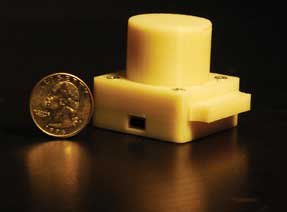
Researchers have described a miniature lensless imaging device that
can be installed on a camera cell phone, which both facilitates imaging and transmits
the raw data to a central computer for processing. Thus, the device could be used
for a host of telemedicine applications involving imaging of blood smear samples,
for instance. Courtesy of Ozcan Research Group.
The system does not provide for physical magnification. “All
the microscopic image reconstruction occurs in the digital domain,” Ozcan
said, “yielding a numerical aperture of approximately 0.2, which is sufficient
to achieve subcellular resolution for imaging of, for example, blood smear samples.”
Because of the unique features of the hologram recording geometry, including the
use of an incoherent source from a large aperture, achieving a decent resolution
and image quality without any artifacts was paramount in the digital reconstruction
process. The researchers addressed this challenge by developing a customized holographic
reconstruction algorithm that iteratively cleans the reconstructed images from artifacts
by recovering the lost phase information.
The cell phone serves a dual purpose in the device’s scheme.
In addition to enabling the actual acquisition of images, it facilitates wireless
transmission of the raw images and related information – demographic data
of the patient, for example – to a central computer at a clinic or hospital.
This reduces the computational burden on the cell phone hardware and provides for
near-instantaneous (<1 s) digital image reconstruction using a GPU installed
on the central computer.
The lensless imaging device is itself both compact and lightweight.
The entire unit, including the battery, LED, sample tray and other mechanical components,
weighs only 38 g.
Ozcan noted that the device’s most important applications
include screening water resources and diagnosing infectious diseases such as malaria,
AIDS and tuberculosis. In the Lab on a Chip study, he and colleagues demonstrated
it by imaging several microparticles as well as red and white blood cells and platelets.
They also measured the waterborne parasite Giardia lamblia because of its implications
for global health. The results from the lensless imaging device were confirmed using
a conventional lens-based microscope, with decent matches in all cases.
The researchers continue to develop the technology for telemedicine
applications. They plan to conduct trials – for diagnosis of malaria, for
example – to test the device in the field. In addition, they are seeking to
enhance its performance by increasing its spatial resolution and extending the technique
to encompass fluorescence imaging.
Published: September 2010