Researchers from multiple institutions collaborated to develop a way to translate information from the mid-infrared (MIR) region, where chemical signatures are most distinct, to the near-infrared (NIR), where existing camera technology is most sensitive. Their new, wide field-of-view system can capture MIR spectral images of fast events or dynamic processes that take place in a matter of milliseconds. The technology could be used to investigate the chemical signatures of cancer and other diseases in ways that would increase accuracy and speed diagnoses.
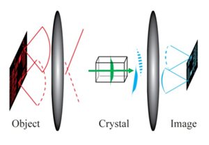
Researchers developed a new system that uses frequency conversion to shift an entire mid-infrared image into the near-infrared wavelength range while preserving the spatial information. The system could be used to look for the chemical-specific signatures of cancer and other diseases. Courtesy of Peter Tidemand-Lichtenberg, DTU Fotonik.
Researchers from DTU Fotonik used nonlinear frequency conversion, a process in which energy is added to a photon to change its wavelength, to develop a detection system that could shift an entire MIR image into the NIR wavelength range while preserving all of its spatial information. Researchers from the Institute of Photonics Sciences (ICFO) developed a new MIR light source that was incorporated into the system. This single-wavelength light source can be tuned to different wavelengths. To generate the MIR light, the light source uses frequency conversion, which allowed the researchers to use the same pulsed NIR laser both to generate the tunable MIR light and to achieve the image upconversion.
A standard CCD camera, in concurrence with the crystal rotation of the upconversion system, acquires the upconverted MIR images containing 64 kpixels in only 2.5 milliseconds, eliminating the need for postprocessing.
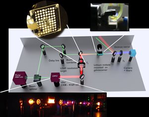
The system incorporates a single-wavelength light source that uses frequency conversion to generate mid-infrared light and can be tuned to different wavelengths. The researchers used the same pulsed near-infrared laser to generate the tunable mid-infrared light and to achieve the image conversion. They demonstrated the system by imaging a gas flow and distinguishing cancerous and normal samples of esophageal tissue. Courtesy of Chaitanya Kumar, ICFO, and Saher Junaid, DTU Fotonik.
“This approach yields high peak-power pulses in perfect synchronism, eliminating the need for sophisticated temporal control of the pulses, leading to images with a good signal-to-noise ratio,” said professor Peter Tidemand-Lichtenberg. “In addition, our optical setup is designed in a way that requires very little post-processing after the images are acquired.”
The researchers demonstrated the imaging speed of their MIR upconversion spectroscopy approach by tuning the illumination laser to match the peak absorption of a gas flow and acquiring a video with 40 images per second. Team members from Exeter University headed a pilot study in which the system was used to evaluate cancerous and healthy esophageal tissue samples. They found that morphology and spectral classification using the new system matched well with the standard stained histopathology images.
Tidemand-Lichtenberg said that the new approach is generic in nature and “constitutes a major simplification in realizing video-frame-rate mid-infrared monochromatic imaging.” The researchers believe that the spectral information provided by their technique could be combined with machine learning to enable faster and possibly more objective medical diagnostics based on chemical signatures, without the need for staining.
The research was published in Optica, a publication of OSA, The Optical Society (https://doi.org/10.1364/OPTICA.6.000702).