Photoacoustic tomography enhances scanning of bodily tissue and its function, resulting in more precise disease detection.
HANK HOGAN, CONTRIBUTING EDITOR
Photoacoustic imaging, or the use of light to create sound, can result in better diagnoses of solid tumors in breast, thyroid, and other cancers, as well as reveal the development of Crohn’s disease, vascular disease, and other conditions. And now, after decades of research and development, the first photoacoustic devices for clinical diagnostics are awaiting FDA clearance.
Photoacoustic (or optoacoustic) tomography can image blood flow, volume, and oxygen saturation, providing important functional information to complement structural data, including the location and shape of specific tissues such as bones or organs. Advancements in sources, detectors, software, and systems are resulting in a reduction of costs and the improved performance of imaging systems.
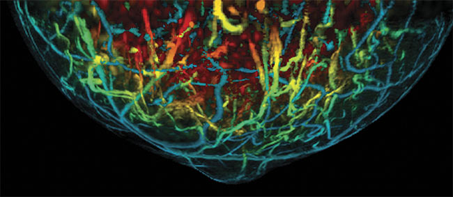
Breast vasculature imaged with photoacoustic tomography. The technique can capture blood distribution and oxygen saturation, indicating the nature of any cancer found. Courtesy of TomoWave.
Analysts predict that the technology is about to take off in the marketplace. In an April 2019 report, QY Research forecast that the global photoacoustic imaging market, which in 2018 reached $49 million, could hit $130 million by 2025 — a compound annual growth rate of 15.3%.
The capabilities of photoacoustic imaging are driving its growth. Lasers or LEDs send a pulse of light into tissue, which absorbs the energy, heats up slightly, and expands. The expansion creates sound, which travels back out of the sample. Transducers pick up the signal, thereby enabling the ability to locate the sound’s origin.
How the imaging works
“[Optoacoustic tomography] allows us to obtain high-resolution, high-contrast images of deep tissues with molecular specificity,” said Alexander Oraevsky, a pioneer in the field and CEO of TomoWave Laboratories. The Houston-based company is commercializing the technology with the goal of developing better breast cancer detection and other applications.
Because molecules have various optical properties, the wavelength of the light pulse can create molecular contrast. Hemoglobin and oxyhemoglobin (the oxygen-carrying form of hemoglobin that makes blood red), for example, are two naturally present molecules that can be imaged. Aggressive cancers need a blood supply to grow. The ability to measure blood distribution and oxygen saturation
deep within tissue without artificial contrast agents gives photoacoustic techniques important clinical capability.
For breast cancer, the ability to distinguish between benign and aggressive lesions is vital. Today, however, 70% to 80% of biopsies performed because of
x-ray mammography and ultrasound re-sults ultimately show no cancer, according to Oraevsky.
“It’s overdiagnosing that causes a lot of financial stress and anxiety,” he said of current screening methods.
Technologies on the market
TomoWave offers products that can be used for preclinical studies on mice and other small animals. The company is working to develop a breast imaging system for clinical use.
San Antonio-based Seno Medical Instruments Inc. is also working with the FDA to get its optoacoustic device on the market. Its first intended application is breast cancer diagnostic imaging, said CEO Tom Umbel. Another application being developed is thyroid imaging. Currently, about 90% of thyroid nodules found by ultrasound imaging are benign, according to the company, and a definitive diagnosis early on could prevent unnecessary biopsies and relieve stress on doctors and their patients.
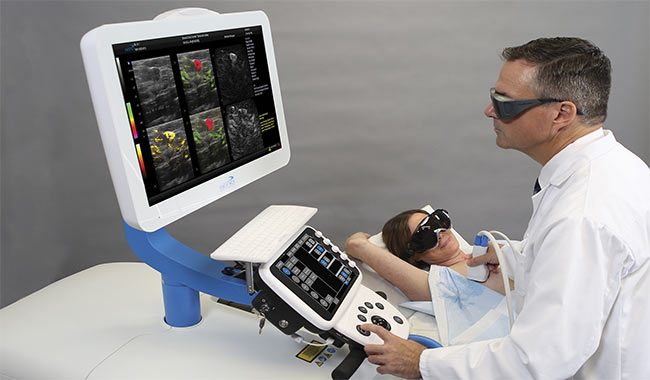
Breast cancer diagnostic imaging could be improved by photoacoustic tomography, with devices currently in development for clinical use. Courtesy of Seno Medical.
Beyond improving diagnostics and reducing needless biopsies, the imaging
technology could be used to gauge cancer treatment. Photoacoustic imaging of microvasculature can spot angiogenesis, the formation of new blood vessels. Angiogenesis plays an important role in the growth of cancer because solid tumors need a blood supply to grow. If chemotherapy is effective, angiogenesis should stop and tissue should die — events that light-sound imaging could track.
“We think this technology has the potential to assess the impact of chemotherapy earlier in the process than we do right now, which is about an eight- or 12-week wait period,” Umbel said.
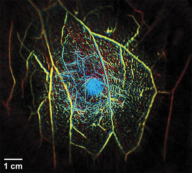
An in vivo 3D photoacoustic tomography image of a human breast. Researchers recently demonstrated the ability to image the entire breast in a single breath (about 15 s). The method could result in the clearest images yet. Adapted with permission from Reference 2.
Steve Miller, Seno’s senior vice president of engineering, said the company has devoted significant resources to adapting laser sources for its medical systems. The sources operate in the near-IR at two proprietary wavelengths. The detectors are normal ultrasound transducers.
The technology at work
A hot topic in the industry is the fusion of imaging modalities — for example, using the same transducer for both ultrasound and photoacoustic images or merging data in the software. If the same detection hardware is used, overlaying the two data sets is easier because the patient doesn’t have to be moved.
Ultrasound provides structural or morphological information about the disease, Miller said, which in its progression can alter tissue. Photoacoustics supplies functional and physiological information, such as angiogenesis and blood oxygenation changes.
“[Photoacoustics is] synergistic from a diagnostic standpoint as well as an instrument structural and design standpoint,” he said.
Looking to the future and to cost savings, one option is to replace the lasers typically used in photoacoustics with LEDs, which would lower the cost of the system significantly. But, currently, the pulses produced by LEDs are too low energy, Miller said. Energy is tied to penetration depth, with a lower energy level leading to shallower imaging.
Despite the present drawbacks of the LED systems, Cyberdyne Inc. of Tsukuba, Japan, is working on such an approach and offers its products to institutions for research purposes. According to a company spokesperson, the goal for developing the LED technology is to “turn it into a medical device that could be used for diagnostic imaging.”
However, the best use of photoacoustic LED systems may be in unconventional diagnostic imaging, according to some investigators. Researcher Ali Hariri and colleagues from the University of California, San Diego said in a March 2018 paper1 published in Photoacoustics that LED-based wearable photoacoustic devices may be useful in therapeutic drug monitoring applications that work by looking at the concentration of a drug in the blood.
While LEDs are used in Cyberdyne’s systems, all other photoacoustic imaging systems offered today use lasers. Munich-based iThera Medical GmbH uses a wavelength tunable laser for a light source, said CEO Christian Wiest, adding that users can opt to image with only a single wavelength. The deciding factor is the optical properties of the tissue and molecules being imaged.
“You choose the wavelength according to what you want to look at,” Wiest said.
What resolution enables
Early in the last decade, iThera began offering preclinical, small animal imaging systems and has since moved on to equipment intended for clinical use. The company’s systems have been used to image Crohn’s disease, among other purposes. For Crohn’s, breast cancer, and other bulk tissue disorders, penetration depth of the imager is important because the affected tissue can lie well below the surface. For imaging of the skin and microvasculature, however, resolution down to as small as 10 µm is more important.
According to Wiest, a trade-off exists between the resolution and penetration depth of imaging, which affects acoustic transducer specifications. The ability to pick up frequencies up to 100 MHz improves resolution but worsens imaging depth, compared to what is achievable with a transducer that can capture frequencies to only 5 MHz. Capturing tenfold-higher frequencies leads to a proportional increase in resolution and, at the same time, a comparable decrease in penetration depth. Wiest said iThera offers a new system that uses ultrawide-band transducers sensitive to signals from 10 to 100 MHz, which therefore enables higher resolution than devices that use detectors in macroscopic imaging.
Wiest also noted that the company’s products are being used in novel ways, such as for quantifying collagen in muscle. This capability enables tracking of the progression of certain muscular diseases and allows assessment of the impact of disease treatments that can be expensive but not necessarily effective.
Regarding the use of these therapies in medicine, Wiest said, “Due to their immense cost, it’s crucial to understand whether they have an effect or not.”
While most photoacoustic products for clinical use are in the approval process, basic R&D continues. Lihong Wang, a medical and electrical engineering professor at Caltech in Pasadena, Calif., heads a group that recently published several papers on related subjects.

Thyroid nodules and cancers can be imaged with photoacoustic tomography, potentially preventing
unnecessary biopsies. Courtesy of iThera Medical.
One paper details how a photoacoustic system could monitor and control nanorobots in the body, such as in the gastrointestinal tract, for medical treatment2. The technology could track the nanorobots’ movements and show when they were in the correct place, and a laser pulse could then activate the robots to deliver a drug.
A second paper reports on how using a UV-MIR-UV sandwich of three laser pulses could enable spectral fingerprinting in tissue3. The MIR pulse creates a difference in imaging between the first and last pulses, with the shorter wavelength enabling a higher resolution and the longer wavelength probing for important molecular signals.
Elsewhere in their published work, Wang’s research group demonstrates the ability to image the entire breast in three dimensions in a single breath hold (~15 s)4. Many transducers and special techniques are needed to attain the speed necessary to produce this clinically important result, which could lead to clearer images than were previously possible.
“This minimizes motion artifacts altogether,” Wang said. “Breathing can cause substantial motion artifacts.”
Researchers watch closely
Clinicians are actively following these photoacoustics developments, which bodes well for the imaging technology’s future acceptance. Jonathan Russin, an assistant professor of neurosurgery at the University of Southern California in Los Angeles, said he would like to see a portable device with excellent resolution that could image the flow and characteristics of blood without use of ionizing radiation or artificial contrast agents — requirements that photoacoustic imaging could satisfy.
Russin said photoacoustic imaging of the brains of small animals has produced good results, despite their skulls, which present an imaging challenge because of their density. Preliminary clinical work involving photoacoustics in neurosurgery has just begun, he said. “This is poised to be the next major clinical imaging platform because of the characteristics of photoacoustics.”
Steven Harms, a radiologist specializing in breast imaging at the Breast Center of Northwest Arkansas in Fayetteville, Ark., uses all currently available breast cancer detection tools in his practice and is aware of their strengths and weaknesses. He’s also familiar with photoacoustic technology and the results achieved so far. He said he thinks the new imaging approach will save money and improve the quality of care when it is cleared for breast cancer detection and integrated into the health care system.
“We’d implement it tomorrow if it was available,” Harms said. “It would be extremely valuable.”
References
1. A. Hariri et al. (March 2018). The char-acterization of an economic and portable LED-based photoacoustic system to facilitate molecular imaging. Photoacoustics, Vol. 9, pp. 10-20, www.doi.org/10.1016/j.pacs.2017.11.001.
2. Z. Wu et al. (July 24, 2019). A microrobotic system guided by photoacoustic computed tomography for targeted navigation in intestines in vivo. Sci Robot, Vol. 4, Issue 32, www.doi.org/10.1126/scirobotics.aax0613.
3. J. Shi et al. (March 4, 2019). High-resolution high-contrast mid-infrared imaging of
fresh biological samples with ultraviolet-localized photoacoustic microscopy. Presented at SPIE BiOS 2019,www.doi.org/10.1117/12.2515616.
4. L. Lin and L.V. Wang (June 2018). Single-breath-hold photoacoustic computed tomography of the breast. Nat Commun,
Vol. 9, Article no. 2352.