Dr. William Henry, Infiniled
New light sources can be tailored with respect to emitter design, emission profile, beam shape and power consumption, offering significant benefit for the development of new fluorescence and life sciences applications.
The use of fluorescence continues to grow in life sciences applications. For analytical applications, it offers the potential for low detection levels, high specificity and minimal power consumption. In imaging applications, it offers benefits such as high signal-to-noise ratios and the ability to view dynamic processes. The availability of a range of low-cost and high-sensitivity light detectors has further accelerated the adoption of fluorescence techniques. However, a number of significant challenges continue to inhibit further performance enhancement across the broad range of target applications.
Not least of these is the challenge of producing high-quality light to excite the target fluorophores. Although initial fluorescence systems used arc lamp or gas laser sources, the size, lifetime and drive conditions of such devices made them troublesome for a range of uses. Semiconductor light sources such as diode lasers and LEDs have mainly replaced these older lamp components, but challenges remain.
A new technology, called microLED, combines the benefits of lasers and LEDs to offer enhanced optical performance of fluorescence-based systems by simplifying the optical path, controlling the excitation spectrum and minimizing the wasted light, and enhancing system performance through reduced power consumption and system size.
Fluorescence techniques are based around three core components: the light source, the fluorophore and the light detector. The light source produces excitation light that is absorbed by the fluorophore. The fluorophore absorbs the excitation light and re-emits it at a different – generally lower – wavelength. This change in the wavelength is known as a Stokes shift and is characteristic of the fluorophore. Finally, a detector measures light re-emitted from the fluorophore. The wavelength, intensity and time required for the re-emission of light can be greatly affected by the ambient conditions of the fluorophore.
Fluorophores can be tailored to selectively bind to a range of analytes and are then referred to as fluorescent probes. When these probes encounter their target, the resulting binding causes a change in fluorescence generated. These changes can be used to detect a wide range of analytes. Fluorescence analytical techniques offer a range of benefits over detection methods: They are highly sensitive, with detection limits of 1 part per trillion. Fluorescence is a noncontact technique, removing the issue of probe fouling and destruction. The binding of probes is reversible – which means that fluorescence techniques can be used to monitor dynamic systems. The technique is also both selective and versatile; it can be applied to a range of target molecules, and the occurrence of false results is low.
Fluorescence techniques have been extended to imaging. In these applications, the image is built up using light generated by fluorescence rather than via reflection and absorption, as in standard microscopy. This fast-growing technique couples the benefits of the fluorescence, such as sensitivity and selectivity, with the ability to view the location of the fluorescence. This is of particular relevance in cellular imaging applications.
A wide array of light sources is used in fluorescence applications – ranging from LEDs to complex gas lasers. The availability of new light source components has resulted in significant progress in the form factor of fluorescence systems, which have evolved from large laboratory-based systems to benchtop and even handheld devices. A goal for fluorescence analytical systems is to produce portable or remotely deployable devices that can be used for quick, efficient detection of a range of targets at the site of sampling. This may vary from the bedside in a hospital to a river where pollutant levels are monitored.
Although initial fluorescence studies relied on broadband light sources and wavelength filtering, the advent of reliable narrowband sources highlighted the true capability of the technique. After the emergence of the semiconductor laser source in the 1960s, these devices began to replace gas lasers in a number of applications. The initial semiconductor sources, based on gallium arsenide materials, were available only at red-to-infrared wavelengths. It was not until the development of UV-to-green semiconductor sources based on gallium nitride materials that sources became applicable to a broad range of fluorescence applications. These devices first appeared in the mid-1990s in the form of LEDs.
Semiconductor lasers now are available in the UV and blue wavelengths. The semiconductor sources have a large number of benefits over both gas and traditional solid-state sources: They are smaller, more robust and cheaper to produce. Individually, LED and laser sources have unique advantages: LEDs are simple to package and control and are available in a broad range of wavelengths; lasers can produce very high light intensity and have controlled emission beams and a small active area. Unfortunately, some drawbacks remain for the solid-state sources. The wavelengths available from lasers are limited, and the emitters require certain conditions (threshold current
and device temperature control) before light is efficiently produced. On the other hand, LEDs have a diffuse emission beam, with light escaping through all six surfaces, resulting in a lower overall intensity.
The microLED leverages the benefits of both lasers and LEDs in a compact package. Similar to a semiconductor laser, this device can produce a quasi-collimated emission beam directly from the chip and extract all the light generated through a single surface. This results in ultrahigh light intensities of >300 W/cm2. As with simple LEDs, these devices do not require high threshold currents or complex heat sinking to ensure stable performance. They also provide power that is linearly scalable with current, allowing the light output and power consumption to be optimized for the requirements of the system.
The new device is fabricated using LED-type material and is available in a wide range of wavelengths, from the UV to the IR. It can generate light at very low current levels, and only nanoamps of drive current are required to generate light visible to the human eye. The light source enables portable or disposable fluorescence applications such as those powered by batteries and is fabricated by the integration of a parabolic structure directly around the site of light generation (Figure 1); the reflector is etched directly into the gallium nitride material and placed at the closest possible position to the light-generating layer, resulting in control of the emitted light directly at the source.
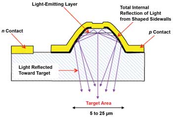
Figure 1. The microLED is made from LED-type material and offers wavelengths from the ultraviolet to the infrared. Fabrication involves integration of a parabolic reflector around the site of light generation; this reflector is etched directly into the gallium nitride material and placed at the closest possible position to the light-generating layer. Images courtesy of InfiniLED.
A standard single microLED pixel is typically 20 µm in diameter; Figure 2 shows a cluster of 25 emitters next to the head of a needle. Devices can be fabricated as single pixels, large clusters – creating collimated emitters up to 1 mm in diameter – or arrays of individually addressable emitters. A single pixel can generate up to 1 mW, and higher powers can be achieved by using clusters. Arrays are available in both 1-D (i.e., linear) and 2-D addressable formats.
The device generates collimated light directly from the chip, enabling a number of benefits for fluorescence applications. It allows for the use of a simplified optics system (i.e., removing the need for a lens), which reduces the size and weight of the final system. The cost of the optical system also is an issue in portable fluorescence applications – especially because lower-cost plastic optical components may produce fluorescence themselves, affecting the accuracy of the system. The diffuse emission generated by standard LEDs reduces the amount of light reaching the sample; this wasted light reduces the efficiency and lifetime of the system. Any stray light that reaches the detector without encountering the fluorophore is also a source of noise in the system.

Figure 2. A cluster of 25 microLED emitters beside the head of a needle. Inset: Close-up
showing parabolic reflector structure.
To further simplify the fluorescence optical system, additional optical components can be integrated onto the new light source, including lenses, filters and polarizers. Optical components require a narrow range of incident angles for optimum performance. The light generated by the new source is within a narrow set of angles and does not require lens or collimating optics between the light source and the active optical components (filters, polarizers, etc.).
The integration of a filter is particularly relevant for fluorescence applications because of the presence of emission tails in all LEDs. Although the full width half maximum of an LED device is ±20 nm, a proportion of light is still generated at longer wavelengths. This is known as the tail of the emission, and it can overlap with the emitted light from the fluorophore, producing false results and reducing sensitivity. Integrating a bandpass filter at the site of light generation – i.e., directly onto the microLED emission surface – overcomes this issue. The filter allows the light required to stimulate the fluorophore through, but it blocks any light at the detection wavelength.
In contrast to LEDs, laser sources have narrow beam angles. However, these devices often require high drive currents before lasing is achieved. The intense light output for the laser results in photobleaching of the fluorophore and necessitates the use of neutral density filters or spreading optics to reduce the intensity of light that reaches the sample. A number of the most common flurophores have been developed based on the wavelengths generated by solid-state and gas lasers: 405, 457, 488 and 532 nm. Semiconductor lasers at these wavelengths are only now becoming available, and their costs can be as high as several thousand dollars per unit.
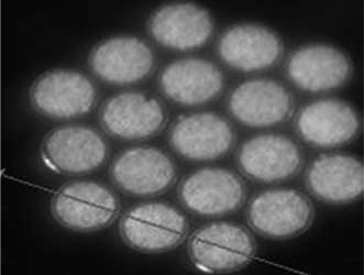
Figure 3. Light from the microLEDs is imaged as it comes through the bottom surface
of the device.
The switching speed of a light source is closely linked to its capacitance, so it’s an active area. In the microLED, the light-emitting area is as small as 50 µm2, which allows the device to be switched at high speeds. In data-transmission experiments, a switching speed up to 1 GHz has been observed during experiments at the Tyndall National Institute – the highest record switching speed for a green LED light source. This is equivalent to an on/off time of 1 ns, ideal for time-resolved measurements. Where increased powers are required, clusters can be used.
An important issue for fluorescence detection applications is the uniform coverage of the target area with light. The emission from standard light sources (both LEDs and lasers) is Gaussian in nature and results in uneven distribution of intensity across the sample. The inherent control of the microLED emission profile allows production of a top-hat profile across the target area without the need for extensive external optical components, improving the performance of a fluorescence system without large additional cost.
The new devices also have been fabricated as addressable arrays of emitters or lines. They can be used to selectively illuminate a sample with high spatial resolution, acting as both the light source and the imaging engine. This simplified design allows for increased efficiency and reduced device size. In one case, the light source has been integrated directly with a CMOS controller, enabling high pixel densities and power output.
Such array devices have a wide range of life sciences applications, including structured light applications such as the development of confocal microscopes without moving components. The UV array head can be used for multiplexed DNA synthesis applications, which involves the de-protection of DNA for site-specific binding.
Studies have shown that the collimated emission from the microLED results in a 20× improvement in the light throughput of a microscope system compared with standard light sources. LED arrays also have been used in optogenetics applications where light is used to stimulate neurons transfected with a photoactive opsin. A microLED array, with its emission controlled at the site of light generation, can be used in implantable optogenics applications because of the miniature form factor and the fact that no external optics are required.
The new devices have drive characteristics similar to standard LEDs and do not require complex control electronics or heat sinking. Effective operating lifetimes are 50,000 h. These devices are based on LED-type material and benefit from the economies of scale available within the industry.
Meet the author
Dr. William Henry is chief commercial officer at InfiniLED in Cork, Ireland; email: [email protected].