Dr. Lin Chandler, Horiba Jobin Yvon Inc., Fluorescence Div.
Time-resolved microscopy is the ultimate tool for investigating dynamic events in cells and subcellular structures. However, it previously was limited when used for applications that required wavelengths
longer than 370 nm because of microscope optics that did not transmit in the deep-UV.
Scientists must perform experiments in the deep-UV
because some applications, such as intrinsic protein fluorescence studies, involve
fluorescence emission in that region.
A new filter-based confocal system
can measure fluorescence lifetime and intensity directly under the microscope in
the deep-UV and visible range of 240 to 850 nm. It is based on an Olympus BX51 microscope
that was modified to transmit light for both excitation and emission to 240 nm.
The system, called DynaMic, provides
single-photon sensitivity and fast acquisition. It can be used with pulsed laser
diodes — available for 265 to 1130 nm —or with pulsed LEDs. The laser
diodes allow confocal measurements. The standard detector (TBX-04) covers 240 to
850 nm. To maintain maximum light throughput, the excitation light source is directly
coupled into the white light-source port on the microscope, and the detector fits
on a pinhole turret attached to the trinocular head.
The turret in the emission light path
comes with pinholes from 0.1 to 10 mm for various spatial resolutions. Lifetime
acquisition can be completed within seconds or minutes for any fluorescence lifetimes
between 100 ps and 100 μs. With an automated stage, this system can map fluorescence
lifetimes and intensity with variable spatial resolution (>1 nm).
NATA (N-acetyl-L-trytophanamide) has
a known standard lifetime of 2.87 ns.1 The time-correlated single-photon counting
microscope system produced similar results. NATA lifetime was measured using a pulsed
280-nm LED for excitation, a 300-nm dichroic and a TBX-04 detector for emission.
The resulting single exponential decay with fluorescence lifetime of 2.9 ns is very
close to the reported value. The system is suitable for intrinsic protein fluorescence
measurement from biological samples, such as calmodulin, tryptophan and tyrosine,
via microscopes because they emit between 290 and 360 nm, which conventional microscopes
don’t cover.
One potential application of this microscope
system is to measure the fluorescence lifetime of a single protein crystal. This
allows scientists to identify and distinguish protein crystals in structural biology.
Drug development usually involves the growth of individual crystals for x-ray diffraction
analysis. It normally takes a long manual process to find the real protein crystal
for structure analysis among many other nonprotein ones, such as salt crystals.
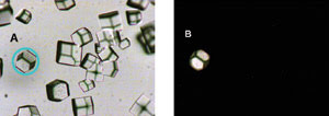
Figure 1. Protein crystals are shown without pinholes in the light path (A). An isolated
single crystal with a pinhole of 0.7 mm is shown in the light path (B).
An automated routine with fluorescence
steady-state measurement has been developed to identify protein crystals.2 A fluorescence
time-resolved measurement could be used to confirm the protein crystal and provide
additional information for its growth and interactions. A single crystal with a
diameter of ~70 μm was isolated using a pinhole of 0.7 mm in the emission
light path and a 10x objective (Figure 1). The fluorescence lifetime from this crystal
was successfully measured with DynaMic under the same conditions as NATA. The results
indicated a triple exponential with lifetimes of 2.1 ns (38 percent), 0.6 ns (8
percent) and 5.5 ns (54 percent). The chi square was 0.96.
The system also can be used for lifetime
and intensity mapping of tissue sections via an automated stage. Lifetime mapping
was obtained from a stained mouse kidney section with excitation from a 455-nm LED,
emission via a 500-nm dichroic filter and the TBX-04. The experiment was performed
using a spot size of 2.5 μm, a 20x objective and a pinhole of 0.1 mm (Figure
2). The mapping of intensity can be obtained at the same time.
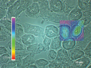
Figure 2. This lifetime map of a stained mouse kidney section was performed using an automated stage.
With time-correlated single-photon
counting’s ultimate sensitivity and UV-VIS (240 to 850 nm) capability, this
microscopic fluorescence lifetime system will find wide applications in biotechnology
and nanotechnology, especially for intrinsic protein fluorescence measurement.
Meet the author
Lin Chandler is senior applications scientist
at Horiba Jobin Yvon Inc.’s Fluorescence Div. in Edison, N.J.; e-mail: [email protected].
References
1. J. Lakowicz, Principles of Fluorescence
Spectroscopy, second edition. P. 283.
2. R.A. Judge et al (January 2005).
An ultraviolet fluorescence-based method for identifying and distinguishing protein
crystals. ACTA CRYST, D61, pp. 60-66.