Whole slide imaging and multiplexing provide rich data extraction and depth of detail while keeping the whole specimen in the field of view.
BRENDAN BRINKMAN, OLYMPUS CORP.
Whole slide imaging is a method principally employed when scientists need to image a large, relatively two-dimensional sample that expands beyond a single microscope field of view. Typical applications include imaging of histological tissues that have been cut into thin slices and placed on a glass slide.
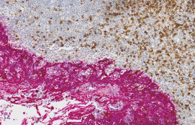
Tonsil CD3 (rm), ImmPRESS Reagent (HRP) Anti-Mouse IgG Immpact DAB (brown), and AE1/AE3 (m) ImmPRESS (AP) (HRP) Anti-Rabbit IgG ImmPACT Vector Red (red). Counterstained with Hematoxylin QS (blue). Courtesy of Vector Labs.
The basic principle of whole slide imaging is to digitally assemble high-magnification images with a small field of view into a larger overview of the sample. Quite simply, the power of this type of imaging, sometimes called digital slide imaging or virtual microscopy, is to give a more complete biological context to a microscopic image. Seeing one or two cells in isolation at high magnification can be informative about a particular cellular mechanism or interaction, but placing that cell in the larger context of whole tissues offers broader avenues of investigation.
It is not just a question of taking a macro image of tissue. The benefit of whole slide imaging to the researcher is its macro-to-micro view, where the microscopic resolution allows greater quantification of images, whether it’s counting nuclei throughout a tissue or tracing axons over large distances in a brain. “Macro to micro” is the typical workflow in which a large overview of the sample is acquired automatically, providing a lower-resolution map to better define micro-level investigation. Because of this powerful combination of resolution and biological context, automated whole slide imaging has become a cornerstone of many research programs. These include everything from archiving serial brain sections to cataloging thousands of samples from a specific cancer type in oncology research, and providing high-throughput image acquisition for serial section reconstruction.

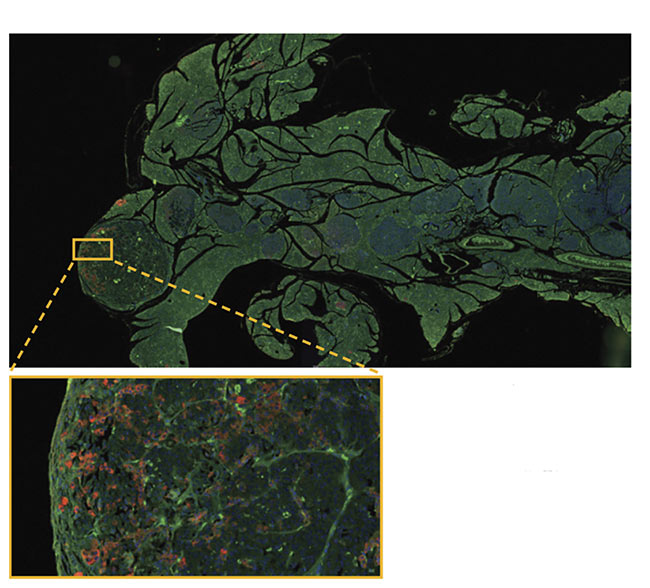
Corticothalamic projection pathways labeled with AAV-GFP and AAV-tdTomato. Courtesy of
Hong Wei Dong/University of Southern California.
For years, these systems were limited to only two imaging modes: bright-field and epifluorescence. In bright-field imaging, transmitted white light passes through the sample, and tissues are visualized through chromogenic stains. But unless you are using stains such as hematoxylin and eosin (H&E), bright-field will not provide any innate contrast. Epifluorescence imaging, on the other hand, enables specific antibody labeling and higher degrees of molecular targeting. It also permits a higher number of stains to be overlaid, which expands the field of multiplexing.
Today, several types of intrinsic contrasts can be used with modern whole slide imaging systems. These sometimes require simple optical systems or filters that enhance contrast in a sample without the need for special stains. The types include:
Phase and polarization. Phase and polarization can be included in whole slide images, providing various levels of innate contrast based on phase ring illumination or polarized light.
Dark-field imaging. Dark-field imaging uses the refractive index differences in membranous structures in tissue. Although some stains can be used to enhance dark-field, the technique can be completely label-free and provide a level of contrast that is comparable to fluorescence. However, this observation mode can be sensitive to dust.
Acquiring a z-stack. It can be taken for granted that whole slide images are only captured in a single plane. But with many modern commercially available whole slide imaging systems, it’s possible to focus through a z-stack, or multiple planes of tissue, either to retain the complete volume of data or a best-focus image. Other benefits include the ability to:
• Acquire multiple planes at
the highest resolution.
• Image samples up to 100 µm thick.
• Image with all available observation
methods.
• Easily obtain information from
all dimensions of the sample.
• Calculate projections (deconvolution,
maximum z, or EFI [extended focal
imaging]).
Oil immersion. For many years, it was not possible to use oil immersion between the lens and the glass slide in a whole slide imaging system to achieve the highest resolution. However, this is now possible thanks to automatic oil dispensing mechanisms, further enhancing the system’s ability to collect high-resolution images.
A combination of imaging modes, or multimode imaging, is most effective when used with the molecular specificity afforded by antibody-labeling techniques. However, there have been complications with labeling efficiently and quantitatively with fluorescence.
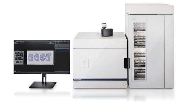
The Olympus SLIDEVIEW VS200 digital slide scanner. Courtesy of Olympus Corp.
Multiplexing in tumor research
Multiplexing has become a useful tool in immuno-oncology research due to the incredibly complex tumor microenvironment. The number of cells, the various tumor invasion methods, and the relationship between the immune system, the tumor, and the different drugs used to combat cancerous diseases are all factors. Compounding the complexity is the number of active substances approved to treat various types of cancers. How does an immuno-oncologist choose the correct drug and identify the right treatment plan?
Multiplexing has been around for some time, but manufacturers have been working to develop a new set of tools that can be more broadly applied and deployed to a variety of tissue locations. Multiplexing is challenging because it requires specialized software, personnel, and equipment to get an assay up and running and applied to a tumor of interest. The multiple-labeling process that is often necessary can require specialized image alignment algorithms, as well as trained staff to use the precision scanning equipment that recognizes tissue and provides images ready for realignment.
Manufacturers have addressed the needs and issues that in the past kept this type of assay from being widely used. To streamline the technology to fit workflow requirements that can ultimately be scaled up, technological must-haves include: high-level multiplexing that addresses target plurality, elucidation of cellular heterogeneity, and sample scarcity; whole slide imaging that captures sample morphology, cellular interactions, and immune infiltration; workflow compatibility that offers high throughput, minimal capital expenditure, and seamless implementation; and software that can identify complex phenotypes with coexpression/colocalization and differential expression.
Images need to be quantified
Particular combinations of optical and staining technologies can lead to the best downstream analysis. Analyzing low-quality images either from a bad reagent set or poor acquisition and optical configuration will not achieve a good quantitative analysis in research. Ultimately, the goal of digital slide scanning is to measure and quantify some aspect of the tissues. However, certain factors need to be considered.
Pancreas stained with DAPI, GFP, and RFP. Courtesy of Rutgers Cancer Institute of New Jersey/ Wenjin Chen.
Transmitted optics. It’s important to have a reproducible and true color representation in H&E staining. Transmitted optics should have spectral characteristics that mimic halogen light sources to reproduce purple, cyan, and pink colors.
Color fidelity and precision. In addition to having the right illumination spectral characteristics, the microscope camera should have excellent color fidelity and precision. A color-corrected camera with the right balance to accurately represent the tissue should be used. Eyeballing digital images can be subjective because different acquisition settings lead to different image outcomes, but color-corrected cameras and ICC (International Color Consortium) profiles replicate excellent color and intensity on computer monitors.
Flatness of field. In a series of images where the field is not flat or where no correction is built in, a halo effect can appear through every field of view, which is a problem for quantification. If this is the case, nonlinear balancing and filtering of every field of view, which takes time and resources, should be used. Consider the optical configuration that will help maximize the flatness of field to lead to a better quantitative result.
Techniques work best combined
Because images can be acquired in volume with a multiple-focus z-stack, a scientist or technician can improve and enhance the images’ contrast using methods such as deconvolution, maximum-intensity projections, or extended focus views. Having an option of methods allows selection of the best focus for the image. Ultimately, the solution to quantitative analysis is a combination of good labeling, good optics, and software that is robust enough for segmentation analysis, garnering statistical information that is biologically relevant.
Meet the author
Brendan Brinkman spent many years working in the stem cell field, including primary mouse neurosphere derivation. After several years as a core imaging facility manager at the University of California, San Diego, he joined Olympus Corp. in 2007 to assist in building novel systems. In 2017, after 2 1/2 years with Olympus Tokyo, where he helped develop next-generation solutions, Brinkman returned to Olympus’ Waltham, Mass., location, where he now works in the Life Science New Business Strategy division as a senior marketing manager.