Researchers at Boston University introduced a multifocus optical microscopy technique for simultaneously acquiring images at differing depths. The method can be added to existing camera-based microscopy techniques, such as fluorescent, phase contrast, and dark-field imaging.
The development will allow users to observe living cells and organisms at high speeds and with high contrast.
Using a z-splitter prism that the research team led by Jerome Mertz assembled from off-the-shelf components, including partial beamsplitters and right-angle prisms, the researchers divided detected light to produce multiple images simultaneously inside the boundaries of a single camera frame. The team built the prism by cementing individual components together with a UV-curing optical adhesive, Sheng Xiao, a member of the research team, told Photonics Media.
Because the parts were of uniform dimensions, the team was able to build its assembly exclusively in its lab.
“One advantage of our z-splitter prism is that it offers different configurations with imaging geometries,” Xiao said. “We can image nine focal planes simultaneously arranged at a 3 × 3 geometry, six focal planes in a 3 × 2 geometry, or thee focal planes in a 3 × 1 geometry. This allows us to take advantage of the rolling shutter readout structure of typical imaging sensors, where we can have higher imaging speed with a more elongated image geometry by cropping the sensor readout area vertically.”
Once the z-splitter prism divides the light, each captured image is focused at a different depth. A camera with a large sensor area and high pixel count allowed the researchers to distribute multiple, high-resolution images on the same sensor without overlap.
Though the number of pixels, low readout noise, and speed of the camera supported successful tests, a camera with a higher number of pixels and higher imaging speed would enable the capture of faster 3D dynamic events, Xiao said, since the system distributes multiple different images on a common sensor. Camera sensors capable of recording at more than 100 fps with 4k or 8k resolution, he said, would be ideal for the technique.
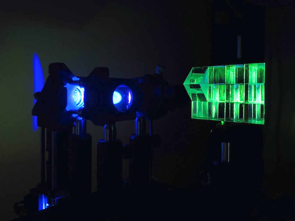
A z-splitter prism (right) produces several images, each focused to a different depth in the sample, in a single camera frame. Courtesy of Sheng Xiao/Boston University.
Compared to using a single image, the multifocal images acquired in the new method made it possible to approximate the out-of-focus background more accurately from a sample. That information allowed the researchers to develop an algorithm for 3D deblurring that successfully eliminated out-of-focus background light.
In wide-field microscopy, background light can distort the quality of captured images.
“Our proposed extended-volume 3D deconvolution algorithm was designed for fluorescent imaging, particularly for imaging thick or densely labeled samples where out-of-focus haze could reduce the in-focus signal contrast,” Xiao said. The process involved extrapolating fluorescent signals beyond the actual imaged volume, and resulted in improving the image contrast and signal-to-noise ratio for imaging applications.
In terms of signal contrast, the method produced an image quality improvement of more than 40× when compared to the raw image. The degree of improvement corresponds directly to the amount of out-of-focus haze present in the sampled raw image: Thicker samples, with more out-of-focus haze, lead to more noticeable improvements with the new technique, Xiao said.
“Optical microscopes, particularly camera-based ones, have been widely used in biomedical research since they allow one to observe biological cells and organisms in their natural state at high speed and resolution,” Xiao said. “For an imaging system, the higher the resolution, the faster an object will be blurred when out of focus. Therefore, for high-resolution imaging of cells and tissues, a sharp in-focus image is only observable over a very thin 2D plane.
“To observe the sample in 3D, one generally needs to translate the microscope focus axially, which inevitably reduces the imaging speed. Our z-splitter prism technique can augment a camera-based microscope for 3D imaging, where objects spanning over an extended volume can be all in focus at the same time, with no moving parts.”
The researchers demonstrated their advance with fluorescent, phase-contrast, and dark-field imaging, and the technique should work in tandem with most camera-based microscopy methods, Xiao said. Incorporating the new deblurring algorithm would allow a biologist to observe 3D, complex biological systems at high speed and contrast with an easy-to-use system.
The researchers imaged various thick samples to demonstrate the capabilities of their technique, including the brain of a mouse. Next steps will involve advancing the technique toward additional imaging modalities to expand biological and biomedical imaging applications.
The research was published in Optica (www.doi.org/10.1364/OPTICA.404678).