Researchers from the University of Tokyo have developed a way to increase the extent and types of information they are able to ascertain about the insides of living cells by using existing microscopy techniques. Their method does not require staining or fluorescent dyes.
To effectively image individual cells, microscope cameras must detect subtle differences in the light passing through parts of the cell, which is itself nearly translucent. Those differences indicate light’s phase. Image sensors are limited by the amount of light phase they can detect. This is referred to as dynamic range.
“To see greater detail using the same image sensor, we must expand the dynamic range so that we can detect smaller phase changes of light,” said Takuro Ideguchi, associate professor at the University of Tokyo Institute for Photon Science and Technology.
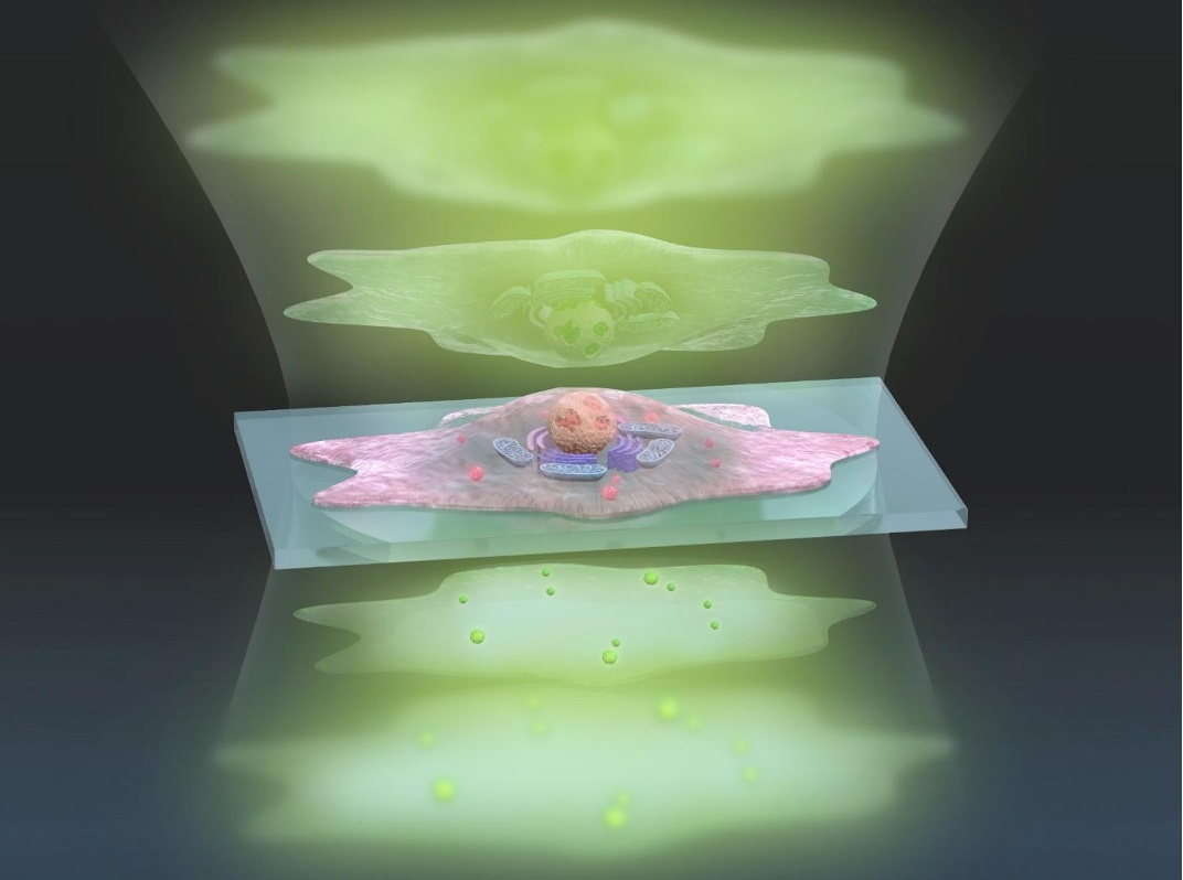
This artistic representation of the ADRIFT-QPI technique shows pulses of sculpted light (green, top) traveling through a cell (center), and exiting (bottom) where changes in the lightwaves can be analyzed and converted into a more detailed image. Courtesy of s-graphics.co.jp, CC BY-NC-ND.
The approach takes into account two exposures, measuring large and small changes in light sensitivity separately. The images are then combined to create a highly detailed final image. The full process is called adaptive dynamic range shift quantitative phase imaging (ADRIFT-QPI).
“Our ADRIFT-QPI method needs no special laser, no special microscope or image sensors; we can use live cells, we don’t need any stains or fluorescence, and there is very little chance of phototoxicity,” Ideguchi said.
Quantitative phase imaging distributes a pulse of a sheet of light toward the cell, and then measures the phase shift of the lightwaves after they’ve passed through the cell. Computer analysis then reconstructs a captured image of the cell’s major internal structures and apparatuses.
Quantitative phase imaging allows researchers to take detailed measurements of unique cells, such as a measurement of the growth of a cell based on lightwave shifts. The quantitative aspect of the technique, however, has low sensitivity, in correspondence to the low saturation capacity of the image sensor. Tracking nanoscale particles in and around cells is therefore not possible with a conventional approach.
The ADRIFT-QPI method overcomes the dynamic range limitations presented by traditional quantitative phase imaging. The final image produced by the combination of the two exposures has 7× greater sensitivity than conventional quantitative phase microscopy images.
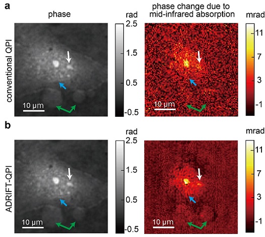
A standard image (top) taken using conventional quantitative phase imaging and a clearer image (bottom) produced using a new ADRIFT-QPI microscopy method developed by a research team at the University of Tokyo. The photos on the left are images of the optical phase, and images on the right show the optical phase change due to the mid-infrared (molecular specific) light absorption mainly by protein. Blue arrows point toward the edge of the nucleus, white arrows point toward the nucleoli (a substructure inside the nucleus), and green arrows point toward other large particles. Courtesy of Toda et al.
The first exposure uses traditional quantitative phase imaging in which a sheet of light is pulsed toward the sample and the phase shifts of the light are measured after it passes through the sample. A computer image analysis program then develops an image of the sample based on the first exposure, and then quickly designs a wavefront of light that mirrors the image of the sample. A separate component, a wavefront-shaping device, finally generates that wavefront with higher intensity light for stronger illumination and pulses it toward the sample for a subsequent exposure.

If the first exposure produces a perfectly accurate image of the sample, the sculpted lightwaves of the second exposure enter the sample at different phases, pass through the sample, and then emerge as a flat sheet of light. It causes a camera to “see” only a dark image.
“This is the interesting thing: We kind of erase the sample’s image. We want to see almost nothing. We cancel out the large structures so that we can see the smaller ones in great detail,” Ideguchi said.
Because the first exposure is imperfect, the sculpted lightwaves emerge with subtle phase deviations. The second exposure shows the tiny light phase differences washed out by larger differences in the first exposure. These remaining differences can be measured with increased sensitivity due to the stronger illumination used in taking the second exposure.
An additional computer analysis reconstructs the final image with expanded dynamic range from the two measurement results.
In proof-of-concept demonstrations, researchers estimate that the ADRIFT-QPI produces images with 7× greater sensitivity than conventional quantitative phase imaging.
According to Ideguchi, the greatest benefit of the new technique is the ability to see tiny particles in context of the living cell without the need for labels or stains.
“For example, small signals from nanoscale particles like viruses or particles moving around inside and outside a cell could be detected, which allows for simultaneous observation of their behavior and the cell’s state,” he said.
The research was published in Light: Science & Applications (www.doi.org/10.1038/s41377-020-00435-z).