Subpixel-shifting digital camera provides artifact-free images.
Brian Graydon, Lumenera Corp.
In cases where cancer is suspected, a pathologist may examine the incisional biopsy collected to determine whether the lesion is benign or malignant and, if malignant, to determine the type of cancer. Hematoxylin and eosin (H&E) staining is one of the most widely used techniques for medical diagnosis and is commonly employed to identify and characterize suspected cases of cancer. Hematoxylin gives basophilic structures a blue-purple hue, and alcohol-based eosin Y colors eosinophilic structures a bright pink.
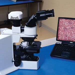
A subpixel-shifting digital camera is used to produce the microscopy image shown on the laptop.
Precise color reproduction, megapixel resolution and high-speed preview are important factors in sample imaging. To ensure a proper diagnosis, it is critical that the colors observed in the microscope match those found in the final image and that the enlarged image is consistent with the original one.
For this application, pathologists can use a microscopy camera such as the InfinityX-32, a 32-megapixel CCD digital scientific camera from Lumenera Corp. of Ottawa (Figure 1). Dedicated microscopy cameras allow the user to view a live preview of the sample, to capture and image it, and to perform any postcapture processing. The camera uses a native 2-megapixel sensor and offers selectable capture resolutions ranging from 2 to 32 megapixels via pixel-shifting technology.
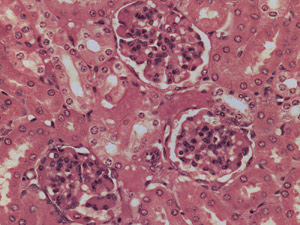
This image of a kidney section stained with hematoxylin and eosin was taken at 40× magnification.
High color fidelity
Traditional color cameras that use a Bayer filter and interpolation to define color but that have no pixel-shifting technology can introduce artifacts into the image and may have difficulty reproducing the exact shade of the H&E stain. Often, the purple-blue hue is rendered as black, and the pinks appear bleached-out. These camera types use a specific arrangement of filters — with each filter containing only one of the three primary colors (red, green or blue) — to create a raw Bayer pattern image. As a result, two-thirds of the color data is missing from each pixel. A complete color image then is obtained by using an algorithm to interpolate the red, green and blue values for each specific point. However, computation errors may occur, resulting in an image whose final color does not accurately match the sample as seen in the microscope’s eyepiece.
In comparison, the sensor in the digital scientific camera undergoes a subpixel shift, moving the sensor across the sample. For precise reproduction, each color filter in the Bayer array is exposed to a specific area of the sample, ensuring that all primary colors are represented in each pixel. The net effect on an H&E-stained sample is that the color in the final image precisely duplicates that seen in the eyepiece.
Variable resolution
A second important function that the camera needs to characterize cancer biopsies properly is the ability to enlarge images for viewing specific areas of interest. With traditional lower-resolution cameras, pixelation occurs when an image is displayed at a larger size because the individual pixels (the smallest piece of information in the image) become visible. It is possible to use anti-aliasing techniques in imaging software packages to smooth this effect; however, this approach can produce blurriness and does not completely remove the pixelation. In a clinical setting, anti-aliasing reduces the sharpness of the detail in enlarged images, resulting in extremely poor presentation.
Besides improved color fidelity, the pixel-shifting technology supplies variable resolution options of 2, 8, 16 and 32 megapixels. During each step of the capture process, the camera shifts the sensor a fraction of a pixel. As a result, objects that are considerably smaller than a pixel may be resolved without pixelation artifacts.
Speed
Previewing video images at high resolution with good color is critical for pathologists when they are examining biopsies suspicious for cancer. With a subpixel-shifting digital camera operating at rates as high as 80 frames per second, that goal is possible.
Meet the author
Brian Graydon is director of business development at Lumenera Corp. in Ottawa; e-mail: [email protected].