From deciphering ancient scrolls to scrutinizing and cleaning must-seen masterpieces, optical analysis uncovers deep secrets — along with best practices for conservation.
MARIE FREEBODY, CONTRIBUTING EDITOR
For hundreds of years, analysis trumped preservation when it came to irreplaceable cultural heritage objects such as paintings, icons, and written works. Today, conservators avoid taking even tiny samples from works of priceless art, making photonics technology an invaluable addition to cultural heritage research.
By delivering technology similar to that used for revealing forged documents or currency, photonics is helping art historians to classify objects or paintings as either genuine and valuable or falsified and worthless. Optical systems are fast, accurate, repeatable, and noninvasive, making them ideal for use on precious paintings, maps, manuscripts, and 3D artifacts.
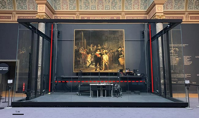
‘The Night Watch’ is surrounded by a glass chamber in the Gallery of Honour at the Rijksmuseum in Amsterdam. The biggest research conservation project in the history of Rembrandt’s masterpiece uses a variety of photonic techniques, including hyperspectral imaging, performed in a specially designed glass chamber so that visitors can watch. See Reference 1. Courtesy of Rijksmuseum, Amsterdam.
For example, John K. Delaney, senior imaging scientist in the conservation division of the National Gallery of Art in Washington, D.C., is part of a team that conducts forensic analysis of paintings and works on paper. Delaney and his colleagues have optimized hyperspectral visible and infrared imaging cameras and image processing to better visualize painted-over compositions and identify and map the distribution of pigments.
“Noninvasive methods allow us to explore the presence of artist materials across the surface of the painting,” he said. “While these methods are not as accurate as microsampling for chemical and material analysis, they allow us to explore more of the artwork.”
Because the hyperspectral cameras can analyze an entire piece of art, rather than a single small point, they deliver a “map” that conservators find easier to understand compared to individual microsample analyses, he said.
The National Gallery of Art team uses molecular luminescence and diffuse reflectance across the 350- to 2500-nm range in both multispectral and hyperspectral imaging. These technologies enable the team to identify pigments and paint binders such as egg tempera, animal skin glue, drying oil, wax, alkyds, and acrylics.
Luminescence imaging spectroscopy highlights materials that emit fluorescence — such as cadmium-based pigments, red lake pigments, or eosin — and fade over time. Today, Delaney’s team and other laboratories are expanding into the MWIR and LWIR to identify and map artist materials that are not easily determined by reflectance or luminescence. These materials include organic paint binders (drying oil, acrylic, alkyd, egg tempera, and gum arabic), paint tube fillers (such as barium sulfate), and some pigments (such as calcium carbonate).
In addition to using the MWIR and SWIR to map materials, the spectral regions of the NIR and SWIR can be used to visualize underdrawings or even previously painted compositions. Typically, the artist begins a painting by drawing out portions of the design with an infrared-absorbing material such as carbon black, applying it directly to an infrared-reflective surface such as gypsum or chalk.

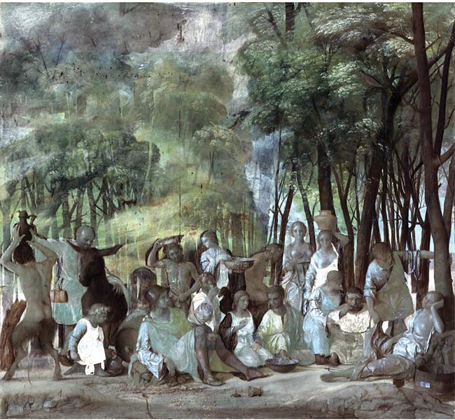
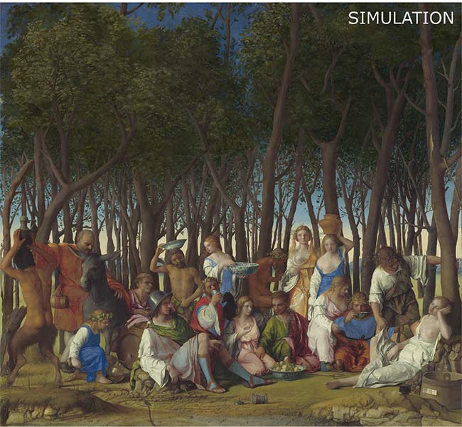
‘The Feast of the Gods’ by Giovanni Bellini and Titian, 1514/1529, from the National Gallery of Art Widener Collection (top). A false-color SWIR image (2175, 1675, and 1325 nm) (center). A simulation of the painting as it appeared after Bellini finished it (bottom). Courtesy of John K. Delaney and Kate Dooley, National Gallery of Art/Becca Goodman, Owner & Paintings Conservator, Goodman LaFon LLC.
“The paint layers are thin enough (tens of microns) and painted with pigments that are less absorbing in this portion of the infrared. Moreover, the paint binders are also transparent at these wavelengths,” Delaney said.
Delaney said Mie light scattering, which hinders imaging through paint layers, decreases in the NIR and SWIR regions. Pigments scatter visible wavelengths more because the particles are several microns in diameter and their optical index is higher than that of the paint binder. The light scatter of visible wavelengths effectively eliminates the contrast difference between the black drawing and the light background of the support. In the infrared, the decreased scattering allows the imaging light to penetrate the paint layer and maintain the contrast between the drawing and the background.
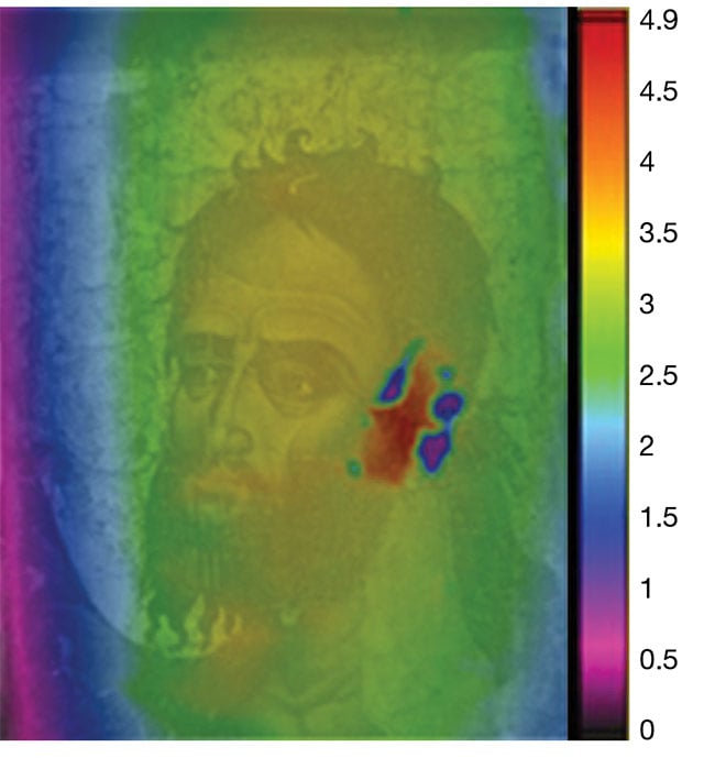
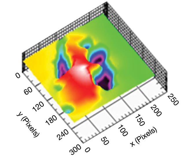
Digital holographic speckle pattern interferometer (DHSPI) full-field scanning reveals a hidden defect in the Byzantine icon of St. John the Baptist at the Byzantine and Christian Museum of Athens in Greece, in work supported by the POLITEIA research project. Courtesy of IESL-FORTH.
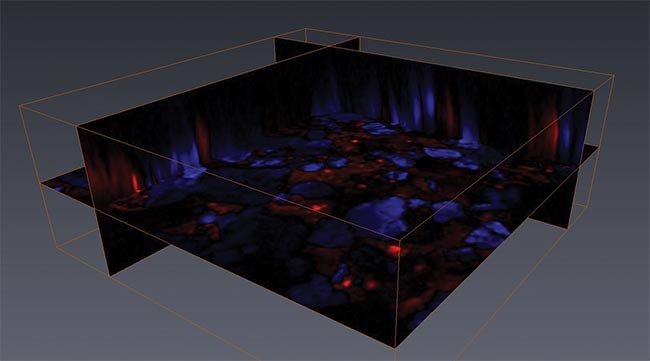
Noninvasive pump-probe volumetric imaging (false color) of a mockup mixture of quinacridone red and ultramarine blue, demonstrating the 3D capabilities of the
technique. See Reference 3. Courtesy of Martin Fischer.
“All these factors allow us to visualize the original drawing,” Delaney said. “Similarly, on a canvas that has been reused, it is possible often to reveal the earlier paintings.”
Wider spectrum, more detail
Until 20 years ago, NIR film and older infrared-sensitive vidicon were commonly used to look for underdrawings. Improvements in digital NIR and SWIR infrared imagers have made such cameras easier to use. At the same time, a shift from multispectral digital imaging into hyperspectral imaging — regularly used in agriculture, defense, and biomedical imaging applications — is beginning to reveal previously unseen markings with more detail than ever before (see sidebar, “Solving Mysteries with More Wavelengths”).
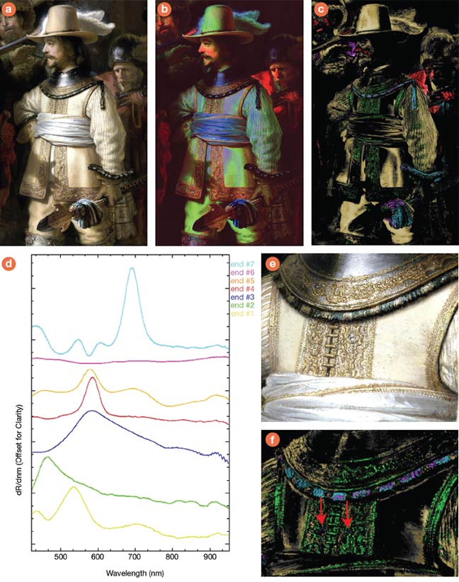
A color image of the figure of Lieutenant Van Ruytenburch (a), a first derivative false-color image of the visible to near-infrared (VNIR) cube, with 620-, 580-, and 520-nm bands used for the RGB channels (b), and a spectral angle map (c) identify the various classes in the image, based on a calculation of the spectral angle for the seven reflectance endmembers (d). A color image of embroidery (e) and a spectral angle map (f) of the same detail, indicating the presence of realgar, an arsenic ore, which the human eye cannot distinguish from similar materials. See Reference 1. Courtesy of Rijksmuseum, Amsterdam.
While many art historians are familiar with using discrete parts of the UV and near-infrared spectrum to reveal hidden details, hyperspectral imaging systems, such as those produced by Headwall Photonics, deliver more detail about the spectral curve and therefore more accurate and specific analysis.
For example, hyperspectral imaging
allowed art historians to date and
authenticate a painting and to determine its physical condition to aid conservation work. Upon examining the “Baptism of Christ” painting, historians at the Benaki Museum in Athens, Greece, found that the figures “MDLXVI,” indicating the year 1566, had been overpainted. Next to these figures, there was what appeared to be another “I,” which would change the year to 1567.
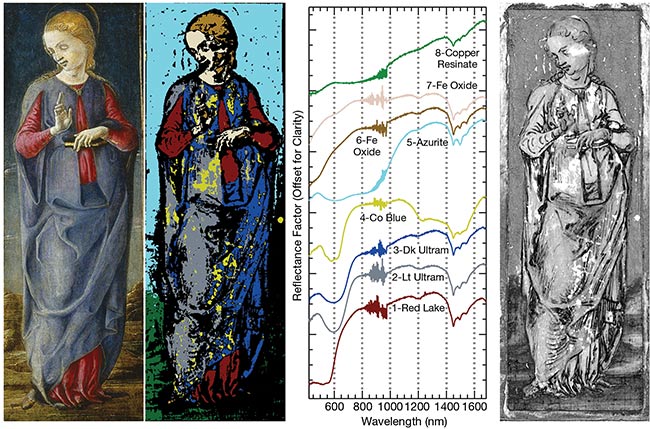
‘The Virgin Annunciate’ by Cosmè Tura, c. 1470/1480, from the National Gallery of Art Samuel H. Kress Collection (left). A spectral angle map of the painting (second from left) with spatial distributions of reflectance spectral endmembers (chart). The hypothesized underdrawing lines obtained from the hyperspectral SWIR image cube (right). See Reference 2. Courtesy of Reference 2.
Given the proximity of the extra “I,” most analysis techniques would have led to an assumption that it was part of the date; however, the chemical information extracted using hyperspectral imaging indicated that the second “I” was part of a nearby scene.
“The advent of digital imaging and specifically narrowband spectral imaging using filters or prisms — or in the case
of Headwall, holographic gratings —
allowed more sophisticated image
processing and analysis,” said Ross Nakatsuji, marketing communications manager at Headwall Photonics.
Today, relatively affordable hyperspectral imaging systems capture details
from the UV (250 nm) to the SWIR
(2500 nm) without requiring the use of consumables such as film and chemicals. Digital storage is also relatively inexpensive. But Nakatsuji said he believes
the industry needs to simplify system
operation and expand image analysis offerings.
Gentle probing with lasers
For Martin Fischer, associate research professor in the Trinity College of Arts and Sciences at Duke University, the bread-and-butter analysis tools are a
standard bright-field microscope (UV
and visible) and a Raman microscope.
Fischer focuses on pump-probe microscopy for molecular imaging in artworks. He joins a growing number of researchers
who apply OCT, photothermal, or nonlinear optical interactions to extract new information from cultural heritage objects.
Pump-probe imaging relies on two trains of moderate-power, very short laser pulses, typically 10−13 s. The first pulse, the pump, excites electrons in pigment molecules or makes atoms within the molecules oscillate. To measure the fate of the excited electron, or the nature of the molecular vibration, a second pulse or probe is sent just after the first.
By changing the delay between the pump and the probe, the researcher can measure the ultrafast dynamics of the molecule. The system response after the pump pulse can be quite specific to a pigment and can be used to determine the pigment’s identity and position.
For example, pump-probe microscopy has been able to nondestructively differentiate and image several red organic dyes, such as carmine, lac, purpurin,
and alizarin. Their similar elemental composition and linear absorption spectra make this differentiation very difficult when using conventional methods, such as x-ray-based or hyperspectral imaging, and without removing some of the
paint for mass spectrometry chemical analysis.
Pump-probe microscopy imaged some pigments in historical paintings deep into paint layers (about 100 μm) while retaining microscopic resolution and molecular specificity. For example, femtosecond pump-probe imaging has been used for pigment separation and nondestructive imaging of the 14th century painting “The Crucifixion” by Puccio Cappana.
“The most exciting developments are the push toward volumetric imaging with increased contrast, multimodal imaging, and portable instrumentation,” Fischer said. “Especially pump-probe imaging techniques, which encompass transient absorption, stimulated Raman scattering, and photothermal contrasts.”
He and his colleagues extract signals with high chemical specificity in subsurface layers of paintings. This information complements data obtained with other powerful analytical tools, such as x-ray-based imaging and spectroscopy, mass spectrometry, hyperspectral imaging, and many others.
“The performance and usability of these techniques has improved so much that some can even be implemented in
the same setup — for example, hyperspectral imaging and x-ray fluorescence (XRF),” Fischer said. “In addition, devices that used to be constrained to the laboratory setting are now finding their way into the field as hand-held or at least portable devices, such as portable XRF or Raman scanners and hyperspectral imagers.”
Although manufacturers and end users agree that the field of art and heritage object analysis is expanding, Fischer said more cross-disciplinary collaborations could benefit both sides.
“It has become increasingly clear, to cultural heritage scientists as well as to researchers in the respective disciplines, that techniques developed for very different applications — for example, in geology, biomedical imaging, materials science, or computer science — can
provide valuable information when
applied to artworks,” Fischer said. “In
exchange, these new applications spur new developments that also can be transferred back into the originating field.”
Options for cleaning and treating
Another technical area that may benefit from cross-disciplinary collaboration is laser cleaning. The technology is used to restore building facades and public sculptures but has benefits for other cultural heritage applications as well. For example, laser cleaning of paintings was introduced in the early 1990s, with obvious noncontact advantages over manual or chemical techniques, but it remains a niche application.
Scientists and conservators continue to study the interactions between radiation and various painting materials, varnish, pigments, and other media. Still, few conservators are trained in laser cleaning, and even seemingly minor tweaks, such as adjusting the laser’s energy level, are critical to its effectiveness.
If the laser wavelengths are strongly absorbed by the material to be removed, and if the pulse duration is short and the fluence values are high, then layer-by-layer material removal is straightforward.
This laser ablation approach, most often achieved using excimer lasers of 248-nm wavelength, selectively removes small amounts of material, such as aged or hardened varnish coatings, overpaints, or soiling. This UV regime is strongly absorbed by the functional groups of the varnishes and of their degradation products.
Lasers emitting in the infrared, such as Nd:YAG and Er:YAG, are strongly absorbed by oxygen-hydrogen (OH) bonds, with a corresponding minimum depth of penetration — a necessary feature when acting on paintings. Unlike ablation, targeting OH bonds is an integrated physical process that is enabled by hydroxylated liquids such as water, alcohol, and mixtures of other liquids.
Because a single-wavelength (1064-nm) Nd:YAG laser interacts very strongly with some materials, but not at all with others, conservators can also clean hard surfaces, such as marble, without damaging the object itself. The energy from the laser is strongly absorbed by dirt, for example, which is ejected and can be subsequently vacuumed away from the object’s surface.
Optical technologies are also helpful for monitoring and assessing the results of a cleaning process. Cleaning of cultural objects is one of the most important aspects of conservation but also the least reversible invasive intervention.
“Cleaning chemically or mechanically, or by laser, without monitoring, and prior to any intervention assessment, are just blind actions,” said Vivi Tornari, senior specialized scientist at the Foundation for Research and Technology Hellas in Greece.
She said monitoring with digital holographic speckle pattern interferometers (DHSPIs) in the lab or museum has proved its value to conservators because the technique can reveal subsurface anomalies, highlighting where consolidation treatments must be applied or how an object reacts to cleaning actions.
Understanding object structure
Structural diagnosis is crucial to preventing the deterioration that can occur inside an object and then spread outward. Such prevention could make damage visualization tools such as DHSPIs important in everyday practice.
DHSPI systems measure the phase variations resulting from surface displacements, deformations of solids, object shape, or variations in refractive index. The systems deliver full-field scanning, rather than point-by-point scanning, which accelerates analysis. The newest systems are also portable and specifically tailored for the cultural heritage field, making them indispensable for curators, Tornari said.
“I think the state-of-the-art now, especially in regard to preventive conservation, is the online DHSPI climate chamber-monitoring workstation, where the research scientist can simulate an environmental condition and monitor the reactions of samples in real time,” she said. “Up to now, we [have had to]
assume physical processes, safe values,
and risk of damage. With a DHSPI
climate chamber-monitoring workstation,
we do not have to assume anymore. Instead we can test any value, any rate of change, any type of fluctuation, from very low and very slow to fast and abrupt, while we record the impact in terms of surface deformation — directly from the object or sample itself.”
DHSPI systems can be combined with simulated infrared thermography to provide insights into the mechanisms of heat diffusion, as well as processes influencing the equilibrium of moisture content that can affect physical deterioration.
‘The most exciting developments are the push toward volumetric imaging with increased contrast, multimodal imaging, and portable instrumentation.’
— Martin Fischer,
associate research professor at Duke University’s Trinity College of Arts and Sciences
One drawback that Tornari pointed out, however, is that automation is still lacking in cultural heritage-tailored technologies, although it is advancing in other disciplines. For example, automating the selection of flaw-indicative fringe patterns could help DHSPIs become a standard conservation tool for structural diagnosis.
Artificial intelligence may underpin future automation efforts, she said. By making the systems easier for end users, requests may in turn increase and boost the market for investors.
From lab to museum
Because the cost of new systems is too high for many buyers, and investors fear an underfunded market, progress is understandably slow.
As an expert with a unique view of both the supply side and that of the end user, Tornari said cost is the biggest
barrier to further commercialization.
Unlike other well-funded specialties such as medicine or defense, funding of
cultural heritage research is severely limited.
“In medicine, most new systems are sponsored directly in the market,” she said. “In cultural heritage, it is the opposite. New systems with many years of successful research and impressive results that would facilitate the conservation and prevent further deterioration are still available only in research laboratories.
William Brown (right), former chief conservator of the Art Conservation Center at the North Carolina Museum of Art (NCMA), and Martin Fischer, associate research professor of chemistry and physics at Duke University, study the painting ‘St. John the Evangelist Reproving the Philosopher Crato’ by Francescuccio di Cecco Ghissi (1375), a gift from the Samuel H. Kress Foundation. In the study, pump-probe microscopy investigates early signs of vermilion degradation, in which the red pigment gradually darkens and turns gray. See Reference 4. Courtesy of Simone Degan/Duke University.
“The conservators and conservation stakeholders seem to be more open to use of new technologies, and requests for services with laser systems have increased; however, a real turn in conservation has not yet happened,” Tornari said.
Headwall’s Nakatsuji agreed and said more “wow” moments, such as work on the Dead Sea Scrolls (see sidebar), will capture the attention of both curators and industry. He said he hopes that current projects, such as Operation Night Watch, will help.
Scientists at the Rijksmuseum in Amsterdam are carefully mapping Rembrandt’s painting “The Night Watch” under the public gaze. Ordinary art lovers can see the science and study the results in real time while the scientists ascertain
the materials that Rembrandt used,
document the painting’s current state,
and create a comprehensive conservation plan.
“The more researchers know about the work and what has been done over the centuries, the safer and more effective the conservation plan will be,” Nakatsuji said. “Helping to popularize this project is a giant glass-enclosed work area where the public can watch as the painting is imaged and treated.”
Better awareness through such public scrutiny has benefits for bringing photonics technology into the public eye, but wider expansion into cultural heritage applications will require a tighter focus on usability and simplification.
Tornari said conservators might
embrace the newest optical methods
better if equipment suppliers offered more educational opportunities, ideally delivered directly at the museums, galleries, and universities that need it. Hands-on experience during these seminars, she said, would give users more confidence with the optical tools, while demonstrating the benefits of optical methods.
References
1. F. Gabrieli et al. (Oct. 15, 2021). Reflectance imaging spectroscopy (RIS) for Operation Night Watch: challenges and achievements of imaging Rembrandt’s masterpiece in the glass chamber at the Rijksmuseum. Sensors, Vol. 21, No. 20, www.doi.org/10.3390/s21206855.
2. K.A. Dooley et al. (Oct. 15, 2014).
Complementary standoff chemical imaging to map and identify artist materials in an early Italian Renaissance panel painting.
Angew Chem Int Ed Engl, www.doi.org/10.1002/anie.201407893.
3. T.E. Villafana et al. (Jan. 21, 2014). Femtosecond pump-probe microscopy generates virtual cross-sections in historic artwork. App Phys Sci, Vol. 111, No. 5, pp. 1708-1713, www.doi.org/10.1073/pnas.1317230111.
4. J. Yu et al. (June 21, 2019). Visualization
of vermilion degradation using pump-probe microscopy. Sci Adv, Vol. 5, No. 6,
www.doi.org/10.1126/sciadv.aaw3136.
Solving Mysteries with More Wavelengths
Spectral imaging can be carried out noninvasively across a wide range of wavelengths. Multispectral imaging goes a step further, enabling the more accurate analysis that has been used to decipher previously
illegible portions of the Dead Sea Scrolls.
Comprising more than 800 scrolls found hidden in cliff caves along the northwestern shore of the Dead Sea, the writings are the oldest known text of
the Hebrew canon, underpinning both Judaism and Christianity.
The manuscripts date to the time of Jesus of Nazareth and are considered a culturally significant discovery. However, 2000 years of aging as well as exposure and even fire damage have made portions of text unreadable to the naked eye.
Although traditional film-based infrared photography reveals hidden characters, the method is limited.
By using a multispectral camera outfitted with a liquid-crystal filter designed to rapidly change wavelengths, a research scientist made never-before-seen text in the scroll visible. Digital images were then fed into a computer with image-processing software to further sharpen the photographs. The additional visible characters provide valuable study material to theologians and historians.
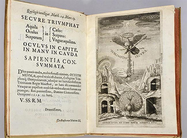
One of the books in the vast collection at Cornell University was Oculus, written by Christoph Scheiner and published in 1619. Optical imaging helped curators determine that their copy had been stolen from the National Library of Sweden; it was later returned. Courtesy of Headwall Photonics/Cornell University.
Hyperspectral imaging goes a step
further and was used to help solve a mystery surrounding one of the most daring thefts carried out at the National Library of Sweden. A treasured book written by the brilliant physicist and astronomer Christoph Scheiner, who developed
theories of optics that later formed the basis for the development of lenses, was stolen from the library sometime between 1995 and 2004 and subsequently sold to an auction house. From there, its whereabouts were unknown until 2012.
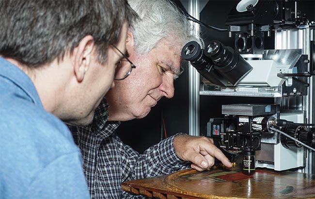
As Sweden worked with Interpol to track down the book, curators at another library thousands of miles away wondered whether their copy might in fact be the stolen book from the Library of Sweden. The head curator at Cornell University Library at the time, David Corson, teamed up with David Bannon, co-founder and managing director of Headwall Photonics, in 2014 to establish the truth.
Headwall’s Hyperspec visible to near-infrared (VNIR) and SWIR sensors were able to yield definitive proof that faint, nonvisible markings on the book were the same size as the bookplates used at the time in the National Library of Sweden’s catalog system, and the purloined book was returned.