
Morgridge Institute to Collaborate with UCSD, Sloan Kettering on NIH 'High-Risk, High-Reward' Project
MADISON, Wis., Oct. 25, 2018 — A project to develop a complete cellular blueprint of zebrafish development, headed by Jan Huisken, medical engineering director at the Morgridge Institute for Research, is one of the 2018 winners of the “High-Risk, High-Reward Research Program” funded by the National Institutes of Health (NIH). The project findings could benefit regenerative biology by precisely defining the role that each cell plays in the full development of a complex organism.
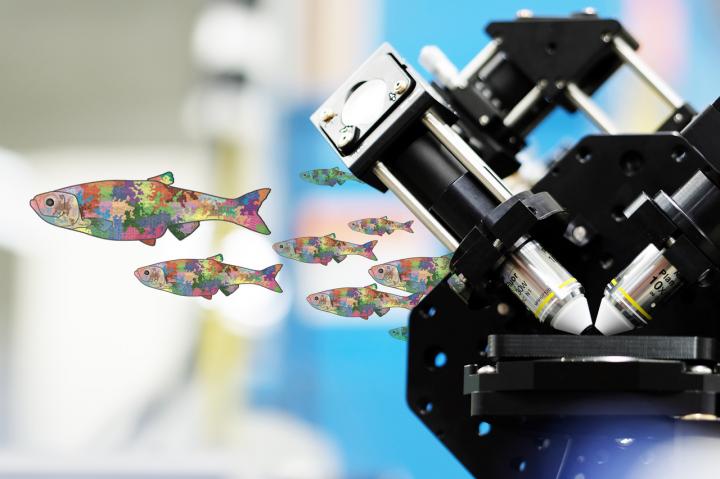
A graphic of mosaic zebrafish paired with the Flamingo light-sheet microscope built by the Huisken Lab at the Morgridge Institute for Research. Courtesy of Jan Huisken, Morgridge Institute for Research.
Three labs will contribute distinct expertise to the effort. Project lead David Traver, a biological science professor at the University of California, San Diego (UCSD), develops unique tools to identify, visualize, and track zebrafish stem cells. Zhirong Bao, a developmental biologist at the Sloan Kettering Institute, provides computational tools to track the movement and function of cells over time. Huisken provides the imaging expertise through light-sheet microscopy, which can noninvasively image living zebrafish embryos for as long as 48 hours.
The project will use laser marking systems to randomly assign a different color to each of hundreds of early-stage cells. Every cell will be a slightly different color than its neighbors, allowing the researchers to track cell migrations.
The team will be using the Huisken Lab’s new microscope, Flamingo. This iteration of a light-sheet microscope is shrunk down to the size of a suitcase and can be shared with biology labs that have fragile specimens that they do not want to transport. The labs at UCSD and Sloan Kettering will be able to use Flamingo to perfect their methodologies, while the high-throughput imaging can be concentrated at Morgridge.
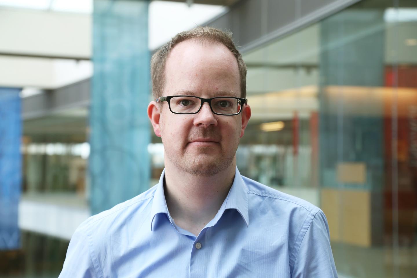
Jan Huisken is the director of medical engineering at the Morgridge Institute for Research. Courtesy of Morgridge Institute for Research.
Huisken said the project’s strongest suit is combining three domains — labeling, imaging, and tracking — to do something no single lab could do alone.
“We want to achieve something in the zebrafish that has only been achievable in much smaller organisms,” Huisken said. “We envision creating an atlas that scientists can look into and see how all of the cell lineages have taken shape.”
Learn more about Flamingo at “Shareable Advanced Light-Sheet Microscopy Using Flamingo,” a free webinar taking place Oct. 25, 1 to 2 p.m. EDT and available for viewing on-demand. The webinar is hosted by Photonics Media, presented by professor Jan Huisken, and sponsored by ASI.
/Buyers_Guide/Applied_Scientific_Instrumentation_Inc/c1021