The speed and versatility of imaging systems enabled by laser components are driving new approaches to life sciences research, with enough detail to dive into complex brain functions.
ERIN DLUGOSZ, MANTAS BUTKUS, AND DARRYL MCCOY, COHERENT
Multiphoton excitation (MPE) microscopy has successfully transitioned from the demonstration of its potential that took place more than 30 years ago to a powerful imaging method used throughout the life sciences. This breadth is reflected in a range of modern techniques, including two-photon (2P) or three-photon (3P) excitation fluorescence microscopy, coherent anti-Stokes Raman scattering, and harmonic generation microscopy. This technology has been applied to a range of applications from research in fields such as neuroscience to analytical assessments of cell vitality for pharmaceutical development and preclinical purposes. And the continued evolution of ultrafast laser systems is helping to bring MPE into new realms.
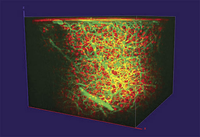
Three-photon imaging of dextran (green) and tdTomato (red) labeled interneurons in a mouse prefrontal cortex, reaching ~1 mm in depth. Courtesy of Timo van Kerkoerle and Marie Guillemant/Neurospin.
The ultrafast laser is the critical component underpinning all MPE microscopy methods. Critical areas of MPE microscopy include in vivo tissue imaging, particularly in neuroscience, where imaging depth, speed, and versatility are important; preclinical and specialized applications for which compact, fixed wavelength lasers can be fully integrated into instrumentation; and emerging applications such as 3P excitation, which require higher power at longer wavelengths.
In vivo imaging
MPE microscopy has historically been used purely as a research method and studies using multiphoton microscopy have long been supported by ultrafast laser oscillators. For many years, these lasers were based on titanium:sapphire (Ti:sapphire) gain medium technology. Neuroscience studies are among the most dominant applications for MPE research, and the demand for longer wavelengths (>1080 nm) to enable deeper imaging of live tissue was a key driver behind the development of femtosecond lasers that combine ytterbium gain medium and optical parametric oscillator technology. This can deliver a much wider, octave-spanning wavelength tuning range (Figure 1). The two laser technologies have their own key advantages in their respective applications.
Laser manufacturers continue to target improvements that enable the capture of brighter images from deeper within the tissue samples at faster frame rates. Certain companies accomplish this by integrating features that address the challenging technology gap between the laser and the microscope. This path to improvement in integration started with group velocity dispersion precompensation. In brief, the accumulated dispersion from microscope elements and other transmissive optics in the laser beam path cause the laser pulses to stretch in time — even deeper tissue samples can add to the total group velocity diversion effect.
However, achieving brighter, higher-contrast images inherently requires shorter pulses with high peak power. So laser manufacturers have incorporated precompensation optics inside the laser systems that pre-stretch the pulses with an opposite amount of group velocity dispersion to fully compensate for all the transmissive optics located in the optical path and microscope itself. This allows for the shortest pulse width at the sample plane. The amount of dispersion precompensation is now just a push-button choice for the user. This feature became even more important as the microscopes are themselves adapted to achieve faster image acquisition or larger field of view.
Controlling the power
The latest integrated feature to improve imaging performance and provide simpler system operation is fast power modulation, down to the microsecond timescale. This is a requirement for optimum performance even in simple raster-scanned experiments, in which laser power should be completely disengaged as the microscope scan head returns the beam spot (fly-back) for the next y scan. Power modulation is even more important for applications scanning large z stacks; as the system focuses deeper into the tissue, natural attenuation dictates the use of higher laser power to maintain constant image intensity. In increasingly sophisticated, high-speed scanning protocols, power control can also be used to maintain the optimum power level relative to the voxel dwell time.
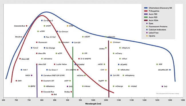
Figure 1. The tuning range of ytterbium-based laser systems can be used for two-photon (2P) excitation of all commonly used probes, fluorescent proteins, and opsins. Courtesy of Coherent.
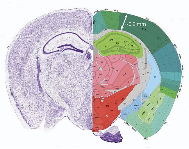
Figure 2. The effective depth limit for two-photon (2P) imaging of the live mouse brain is ~1 mm, thus restricting this method to studies of the cortex. Courtesy of the Mouse Brain Atlas/Allen Brain Institute.
Until recently, power modulation often required the use of external modulators between the laser and the microscope. There are two types of modulator devices that can be used: acousto-optic modulators or electro-optic modulators. Acousto-optic modulators are more reliable and support higher contrast modulation, but electro-optic modulators were often preferred, particularly in home-built solutions because they are much easier to set up. Now laser manufacturers offer this functionality as a seamlessly integrated optional feature within the laser, based on the better performing acousto-optic modulator. This turnkey feature can be called total power control.
Multimodal applications
The growth in multimodal applications and in preclinical settings illustrates just how far MPE microscopy has come in its journey from a new scientific interrogation method to a mainstream imaging technique. Multimodal applications encompass instruments and experiments that combine MPE microscopy with another imaging method. These applications are quite different from other emerging preclinical applications in oncology and other areas of medicine, but they have a common need for advanced laser sources. In contrast to versatile lasers with a full array of features designed for MPE research, these applications can benefit from a compact push-button femtosecond source. This is embodied in lasers such as Coherent’s Axon series that deliver single wavelength output, but which still drive acquisition of high-contrast, high-resolution images due to their very short pulse widths and internal group velocity dispersion precompensation. This minimizes the pulse width at the sample. They also provide an integrated acousto-optic modulator option.
Of course, a key question for single wavelength lasers is: What wavelength(s)? This is determined by closely matching the needs of significant applications within the practical limitations of available laser materials and frequency harmonic/shifting crystals. Lasers with an output at 780 nm provide a great example.
Use of excited fluorescence
There is fast-growing interest in the discipline of microscopy to use incipient fluorescence signals from important metabolites such as reduced nicotinamide adenine dinucleotide (NADH) and flavin adenine dinucleotide (FAD), which can be excited by single-photon absorption in the ultraviolet range. The fluorescence from these molecules can indicate details of cellular vitality, specifically how cells are “burning” sugar to generate energy. Healthy cells generate most of their energy through a process called oxidative phosphorylation, wherein energy is harnessed through a series of protein complexes embedded in the mitochondria. Many cancer cells and otherwise faulty or distressed cells get their energy from a less efficient process called glycolysis, wherein glucose is converted into pyruvate, an acid. The ratio of the amount of FAD relative to NADH is called the redox ratio and is well established as an indicator to reveal which process predominates. This is valuable information in clinical analysis of oncology patients and in drug discovery work.
Early MPE imaging of NADH and FAD used two different wavelengths to match the absorption maxima of these two targets. But this necessitates the high cost of using two femtosecond lasers, or the complexity of a tunable laser. Fortunately, researchers such as Ammasi Periasamy, a professor at the University of Virginia, have since shown that a single intermediate wavelength, at 780 nm, could simultaneously excite with sufficient efficiency to provide the all-important redox ratio1. Therefore, laser manufacturers responded by developing compact, single-wavelength lasers at this specific wavelength.
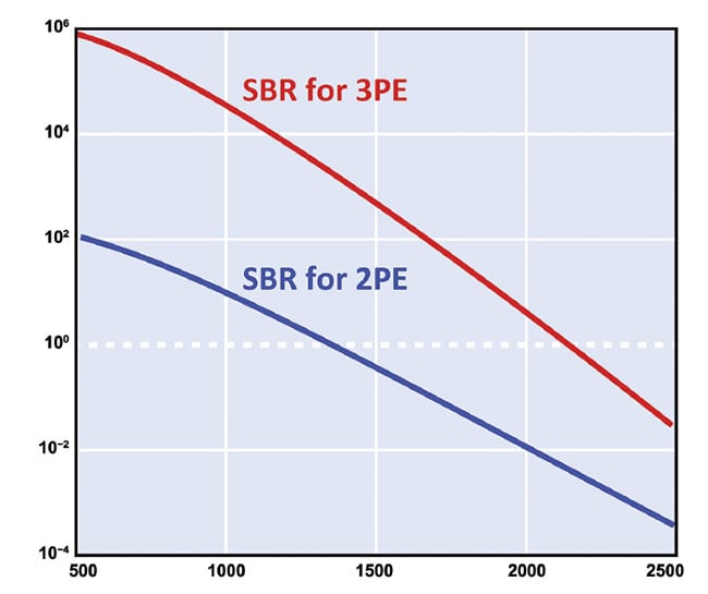
Figure 3. Assuming a scattering length of 250 µm, the signal to background ratio for three-photon (3P) imaging at an excitation wavelength of 1280 nm is up to three orders of magnitude higher than for two-photon excitation (2PE) at that wavelength. As a result, 3P imaging theoretically enables imaging at a depth of 2 mm and beyond and is dependent on the laser power and duty cycle. 3PE: three-photon excitation; SBR: signal to background ratio. Adapted with permission from Reference 2.
For completeness, it should be noted that this requires careful spectral filtering to avoid crosstalk to separately detect the fluorescence from the two species using two photodetectors. But this is a relatively simple and economical accommodation compared to using two ultrafast lasers. The 780-nm wavelength is also well-suited for another label-free imaging technique — namely, second harmonic generation for imaging collagen fibers. In addition to 780-nm wavelengths, lasers are also available at 920-nm and 1064-nm wavelengths, all in the same form, fit, and function covering key popular nonlinear imaging spectral windows.
3P excitation
As previously noted, researchers in neuroscience and intravital imaging are seeking to image deeper into live tissues, which is made possible with the use of longer wavelengths. The maximum imaging depth with 2P imaging depends on the type of tissue and how much signal averaging a researcher is willing to tolerate. It also depends on the choice of excitation wavelength, which is limited by the matches to fluorescent probes and proteins that are available.
In brain tissue, the practical limit for 2P excited fluorescence methods is <1 mm. This limit has become increasingly frustrating for neuroscientists studying the model mouse brain, because the mouse cortex has a total depth of ~0.9 mm, thus limiting 2P imaging to this region (Figure 2). In addition, the area immediately below the cortex (the white matter) has higher scattering, decreasing the scattering length to ~40% of the gray matter that forms the cortex. As a result, imaging beyond the cortex using 2P microscopy is impractical.
However, as long ago as 2013, Professor Chris Xu and colleagues at Cornell University demonstrated that 3P excitation could provide greater depth for brain imaging by using lasers at either ~1300 nm or 1700 nm, which match nicely with the 3P absorption peaks for green fluorescent proteins (GFP) and red fluorescent proteins (RFP)2.
They noted that the 3P excitation mechanism is less efficient than 2P excitation, so 3P will generate much weaker signals. However, they explained that the key imaging metric for deeper imaging is not the raw signal strength, but the signal to background ratio. The higher-order dependence on laser power actually leads to lower out-of-plane excitation for 3P fluorescence when compared to 2P excitation. As a result, they showed that the signal to background ratio can be an incredible three orders of magnitude higher for 3P than 2P in some cases (Figure 3). This supports imaging at a depth of 2 mm and beyond, depending on the laser power and duty cycle (hence pulse energy). An example of 3P imaging is shown in the opening image.
Lasers advance the application
The lower excitation efficiency of 3P requires much higher peak power than for 2P; therefore, the ultrafast lasers developed for 2P could not be used to induce the absorption of three photons in the sample. The high peak power can be reached using a Ti:sapphire oscillator-amplifier setup, but obtaining images in a practical timeframe also dictates the need for a high (>1 MHz) repetition rate and high average power of tens of watts. This combination is simply not a practical option with Ti:sapphire-based lasers systems.
During the past several years, two aspects have changed the environment for 3P excitation. First, an increasing number of researchers have become interested in this technique and its ability to probe beyond the mouse cortex. This has created commercial demand for a suitable laser source. Second, and just as important, amplified ytterbium lasers have been successfully developed. These ytterbium amplifiers can now deliver tens of watts of output and repetition rates up to 50 MHz. By combining one of these amplifiers with a commercially available, tunable optical parametric amplifier and scaling down the repetition rate accordingly (typically in the range of 1 to 4 MHz), laser light in both the 1300-nm and 1700-nm range can be generated with suitable characteristics for 3P imaging. However, there is an inherent level of complexity in this two-box long wavelength generation approach, and a lack of integrated power modulation and dispersion precompensation, which are even more necessary for 3P excitation than for 2P.
To support the widespread use of 3P for imaging of the brain, a one-box laser source should be developed, in which the ytterbium amplifier and optical parametric amplifier are integrated in a compact, simple platform. To aid in the simplification of the technique, Xu’s group has recently shown that while 1300-nm laser light is ideal for exciting GFP, it can also excite RFP and some other longer-wavelength probes with enough signal to background ratio for imaging3. This has enabled laser manufacturers to define one-box single-wavelength 1300-nm sources optimized specifically for use in 3P excitation microscopy.
Expanding use of MPE
To summarize, multiphoton microscopy is a dynamic field characterized by diverse techniques and applications. Its continued growth depends on making it accessible to the broadest possible spectrum of users, rather than only laser specialists. Laser manufacturers will continue to work in close cooperation with cutting-edge users to stay abreast of all key developments and to support these developments with new lasers and innovative laser features.
Meet the authors
Erin Dlugosz is a product line manager at Coherent Corp. She received her doctorate from Temple University, with a research focus on vibrational and rotational Raman spectroscopy; email: erin.[email protected].
Mantas Butkus, Ph.D., joined Coherent in 2013 and is a product line manager for multiple laser products for scientific and industrial markets; email: [email protected].
Darryl McCoy is director of product marketing at Coherent Inc., where he manages ultrafast tunable and fiber laser products for nonlinear microscopy and instrumentation markets; email: [email protected].
References
1. R. Cao et al. (2020). Multiphoton FLIM imaging of NAD(P)H and FAD with one excitation wavelength. J Biomed Opt,
Vol. 25, No. 1, pp. 1-16.
2. N.G. Horton et al. (2013). In vivo three-photon microscopy of subcortical structures within an intact mouse brain. Nat Photonics, Vol. 7, No. 3, pp. 205-209.
3. Y. Hontani et al. (2021). Multicolor three-photon fluorescence imaging with single-wavelength excitation deep in mouse brain. Sci Adv, Vol. 7, No. 12, p. eabf3531.