
Multiphoton Microscopy Technique Advances into Clinical Settings
An instrument developed and introduced by researchers at the University of California, Irvine (UC Irvine) demonstrated the potential to expedite the adaptation of conventional multiphoton microscopy (MPM) for use in clinical settings. The compact, fast large-area multiphoton exoscope, or FLAME instrument, which the researchers described in a published paper last year, addresses the relatively limited field of view (FOV, or scanning area) that is associated with the current MPM technology for clinical skin imaging.
FLAME additionally increases the speed of the signal acquisition time without compromising imaging resolution.
“Rapid noninvasive in vivo and ex vivo multiphoton-based imaging with molecular contrast and high spatial resolution could become an important tool for maximizing diagnostic efficacy and guiding therapy in surgical procedures,” said Mihaela Balu, principal investigator at UCI Health Beckman Laser Institute & Medical Clinic, and senior author of the 2020 paper describing FLAME.
Two-photon microscopy was introduced and subsequently patented by a Cornell University-based team in 1990; the technique involved the application of two-photon excitation of fluorescence to the established laser scanning approach that is used in confocal microscopy. The ability of MPM to generate 3D submicron resolution images of superficial tissues based on endogenous two-photon fluorescence and second harmonic generation signals led to its widespread use in biomedical applications, Balu said.
Subsequently, the potential of MPM for noninvasive imaging of human skin was established with a clinical tomograph (called MPTflex) developed by Jenlab, GmbH.
However, this technology delivers a limited FOV and requires relatively long signal acquisition times.
As a consequence, the technique has struggled to gain traction in clinical settings, despite its ability to provide label-free morphological and metabolic tissue information.
The FLAME system pairs deep learning image reconstruction with an optical and mechanical scanning mechanism, to allow comprehensive tissue inspection in minutes. The computational approaches involve use of content-aware image restoration (CARE), which compensates for a low signal-to-noise ratio at high imaging speeds over multimillimeter scale images.
Compared to the current clinical MPM system, FLAME offers the potential for up to a 16× larger FOV, as well as a 1000× maximum scanned mosaic area at up to 40× greater imaging speeds per unit area, the researchers said.
A time-resolved single-photon counting detection system is also incorporated into the FLAME platform, to further enhance image contrast.
Portability is another area of focus for the UCI team; the standard MPM technology platform typically requires a large ultrafast laser and an optical table setup. The UCI team replaced a Ti:sapphire laser and an associated chiller with a Carmel X-780 fiber laser from Calmar Laser, USA. The fiber laser avoids the requirement of a chiller and features a laser head greater than 70× smaller than that of the previously deployed Ti:sapphire system.
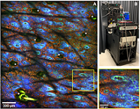
Image acquired in vivo at 60-µm depth from the forearm of a volunteer (A) by using the FLAME imaging platform (B). The macroscopic and the close-up images show collagen (SHG, blue) and elastin (TPEF, green) fibers, pigmented keratinocytes (TPEF, red), and hairs (TPEF,*).The 10-MP macroscopic image (2.5 × 3 mm2) was acquired in ~30 s. Courtesy of UC Irvine and Calmar Laser.
Since the instrument’s introduction, the researchers and their collaborators have further tested it. The FLAME system has been upgraded to integrate a stable patient-imaging head interface, Balu said, which allows in vivo imaging over millimeter-scale areas. Specifically, with automatic data acquisition software, the researchers have gained the ability to obtain real-time macroscopic images over millimeter-scale areas.
Advances to the laser component of the system have further enhanced the imaging signals that are obtained using FLAME. Team members added a pre-chirp capability to the compact head. This compensates for the dispersive effects of the microscope system, which in turn ensures that the shortest pulses impinge on the target area and allows users to acquire the highest imaging signals while the patient is exposed to the lowest laser average power.
The current version of FLAME is being used in a clinical setting to evaluate its potential for noninvasive early diagnosis of skin cancer. In addition to skin cancer diagnoses, the system is being tested for monitoring the effects of therapies on skin morphology and functionality.
The research was published in Scientific Reports (www.doi.org/10.1038/s41598-020-75172-9).
A summary of the research on the FLAME system was presented in the Photonics in Dermatology conference at SPIE (Photonics West) BiOS 2021 (www.doi.org/10.1117/12.2579260).
/Buyers_Guide/Calmar_Laser/c2190
/Buyers_Guide/JenLab_GmbH/c7342