By analyzing blood flow, the structure of the brain, and concentrations of oxyhemoglobin and deoxyhemoglobin, information about when and where an event occurred can be isolated.
IAN READ, SPECTRA-PHYSICS, MKS INSTRUMENTS
Every year, approximately 5.5 million people worldwide lose their lives to stroke. According to the Centers for Disease Control and Prevention, stroke-related costs in the U.S. total nearly $50 billion annually1. Stroke is the fifth leading cause of death and a major cause of serious disability in adults in the U.S. Because of its impact on the health of those stricken and on society as a whole, several health agencies are committed to funding research to enrich our understanding of its causes and effects. This research, which incorporates a number of imaging modalities, is aimed at answering several key questions: What risk factors contribute to the likelihood of stroke? What is the core mechanism that causes it? What determines prognosis? How does the brain recover?
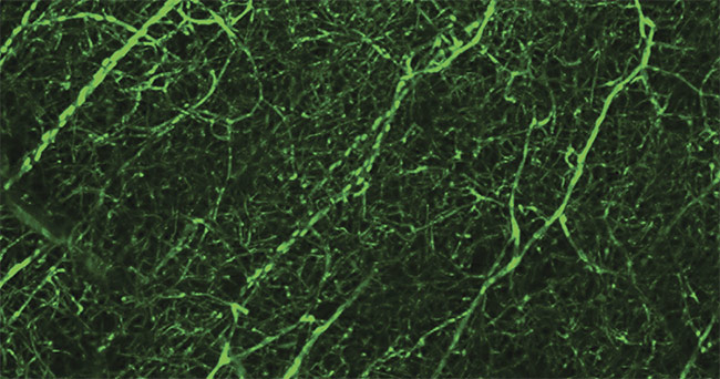
A two-photon image of mouse brain vasculature. Courtesy of Igno Schiessl/Manchester Bioimaging Facility.
To answer these questions, several diagnostic technologies are being used both in the clinical setting and in the laboratory. For example, magnetic resonance imaging (MRI) and computed tomography (CT) are among the most common and most familiar diagnostic tools used in hospitals. Perhaps the most beneficial, albeit less familiar, are the modern optical technologies used in the laboratory, such as near-infrared (NIR) spectroscopy, that can unlock the mysteries of traumatic events such as a stroke.
NIR spectroscopy plays a key role in researchers’ and clinicians’ understanding of this important topic2,3. Its importance is mainly due to the fact that biological tissues are partially transmissive in the NIR spectral region (750 to 2500 nm). Experiments have typically been designed to examine the structure and function of the brain before, during, and after a stroke event. Several key technologies are being used to further advance the understanding of stroke.
What is a stroke?
When a disruption of blood flow occurs to one or more areas of the brain, the event is diagnosed as a stroke. The most common type, ischemic stroke, occurs when the disruption is caused by a blood clot. This type accounts for approximately 90% of all stroke events. If the disruption is caused by a rupture of one or more of the blood vessels, the event is called a hemorrhagic stroke, which is significantly more dangerous. Only 26% of people survive this type, compared to an 80% survival rate in ischemic cases. In addition to being potentially fatal, stroke has been the leading cause of paralysis in adults since 20134.
In general, experiencing a stroke can be catastrophic, resulting in lingering effects and the potential loss of certain physical or psychological functioning. A thorough understanding is therefore imperative for providing information to patients about warning signs and how to incorporate preventative measures and make behavioral changes. If a stroke does occur, the factors that determine the likelihood of recovery need to be established, along with which ones influence an individual’s prognosis. It is also helpful to examine whether a person exhibits particular predictive markers that could determine stroke severity and how much they may be able to recover.
NIR spectroscopy in biological tissue relies on measuring the concentrations of oxyhemoglobin (oxygenated hemoglobin) and deoxyhemoglobin (deoxygenated hemoglobin) (Figure 1). When a stroke occurs, the concentrations of these hemoglobin species can be evaluated to determine whether, when, and where the blockage occurred and which area of the brain may have been affected5.
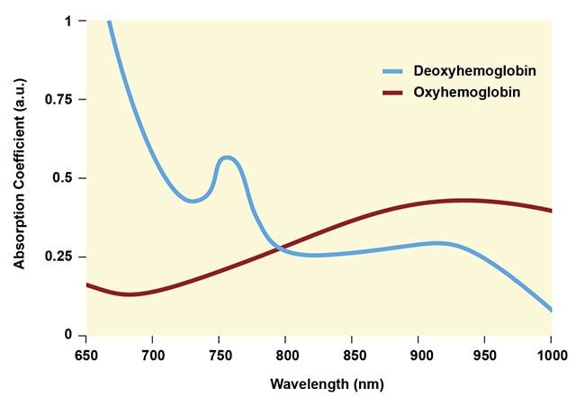
Figure 1. Absorption spectra for oxygenated and deoxygenated hemoglobin. Courtesy of MKS Instruments.
Because brain tissue is heterogeneous, measuring these concentrations accurately is vital to pinpointing problem areas. In general, steady-state absorption is commonly used to determine concentration. Absorption is typically calculated using the Beer-Lambert law,
A(λ) εbCi
where A is the wavelength-dependent absorption, ε is the molar absorptivity, b is the optical path length, and Ci is the concentration of species i. In most applications, the Beer-Lambert law is applied using a standard cuvette with low molar concentrations and no scattering. However, in the case of brain tissue, because of the significant heterogeneity, the optical path length cannot be precisely defined. In addition, because the photons undergo significant scattering, it is difficult to determine which photons are lost due to absorption and which are lost due to scattering. For these reasons, continuous-wave (CW) NIR spectroscopic measurements must be carefully scrutinized to obtain meaningful results.
For example, a typical CW NIR spectroscopy experiment employs a single-wavelength source that is centered on the absorption peak for each species (Figure 1). Several commercial laser systems are available to preferentially excite deoxyhemoglobin (~750 nm) and oxyhemoglobin (~950 nm). The light is fiber-coupled and systematically routed to specific regions of the brain5. In this configuration, the experiment is performed at fixed emitter-detector distances to more clearly determine path length.
Using this configuration, localized differences in oxy- and deoxyhemoglobin ratios can be monitored and evaluated in specific spatial regions across the brain. For stroke patients, this tool provides a real-time measurement of the affected region. The laser apparatus is compact and presents a more practical alternative to MRI at the point of care. However, unlike MRI, correlating CW NIR results to specific events remains challenging. To learn more, time-domain experiments are necessary.
Time-resolved NIR spectroscopy
Determining absolute concentrations of oxy- and deoxyhemoglobin provides more functional information following a stroke event. Recent studies have used time-resolved NIR (TRNIR) spectroscopy to better characterize the event and how oxygen levels are directly influenced before, during, and after the event5. Interpreting this information can inform medical personnel about the severity of the event and the prognosis.
Strokes can affect the brain differently depending on where the blockage occurs along the circulatory network. When the disruption of blood flow happens at a major arterial pathway, this is called large vessel occlusion (LVO) stroke. This type is particularly severe because it causes a significant drop in blood flow to a larger area of the brain. Another type, in which the ischemic blockage occurs at the extremities of the circulatory network, is called a lacunar stroke. In both cases, TRNIR spectroscopy can provide key signatures of oxy- and deoxyhemoglobin concentration that can be correlated to outcome when coupled with MRI and CT images.
Because the NIR apparatus is compact, it can be brought to the patient, and the measurements of the patient’s condition can be monitored more easily than with either MRI or CT. To perform these measurements, the apparatus is similar to that for CW NIR spectroscopy except that a picosecond pulsed laser is used as the excitation source. Experimentally, these measurements are identical to time-correlated single-photon counting. A typical dataset is shown in Figure 2.
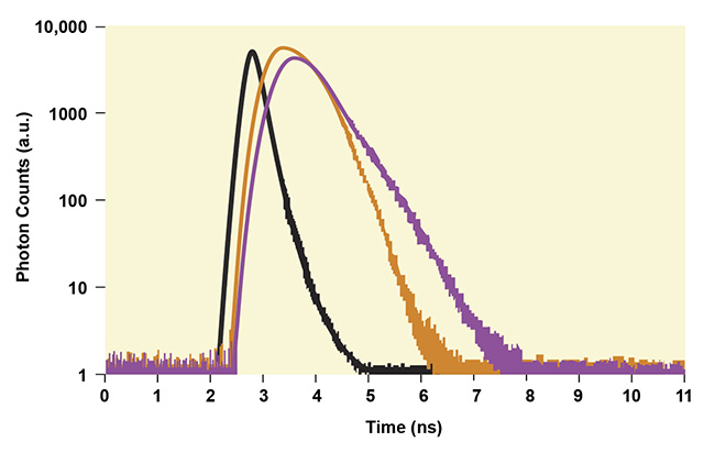
Figure 2. Graphical rendering of typical decay profiles measured with time-resolved NIR
spectroscopy (purple and orange curves). The instrumental response function (black).
Courtesy of MKS Instruments.
A picosecond pulse from a Ti:sapphire or diode laser in the 750- to 950-nm wavelength range is fiber-coupled to the specimen at targeted input locations (Figure 3). A small portion of the incident laser pulse is used to start the gating electronics. As the incident photons propagate through the sample, some will be absorbed and some will scatter. A small percentage of photons will scatter into the detector electronics and stop the timing gate. Several thousand similar cycles will build the histogram shown in Figure 2.
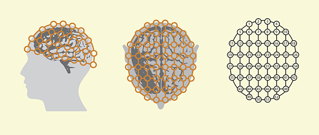
Figure 3. A typical apparatus for measuring time-resolved NIR decay profiles in a clinical setting. Scattered photons are monitored as a function of incident light and detector position. Courtesy of MKS Instruments.
To reduce interference from the incident laser pulse traveling directly to the detector and affecting the desired readout, the intensity is kept low and the number of detected photons is kept small relative to the number of excitation cycles. Time resolution is determined by the laser pulse width and the experimental electronics that are calibrated using a direct line configuration from the emitter to the detector. The direct line measurements provide the impulse response of the experimental apparatus and define the time resolution of the measurement. Figure 2 shows the instrument response function as a black curve.
In general, the probability that a photon will reach the detector is determined by the effective path length, which has contributions from absorption and the scattering efficiency of the sample3,5. In these experiments, the measured decay times are influenced by the presence of a stroke. The decay times are compared in various spatial regions, and conclusions are reached based on the severity of the event. For example, the increased absorption from oxyhemoglobin present in stroke-affected regions will translate as faster decay times. Clinical outcomes are later correlated to this data, which provides valuable information on which to build future clinical applications of NIR spectroscopy for health care settings.
Current research
The clinical studies outlined above provide valuable information regarding stroke outcome. They help physicians to monitor patients in real time during a stroke and its subsequent treatment, and throughout the recovery process. However, NIR spectroscopy using femtosecond (10−15-sec) pulses is being used to reveal even more detailed information. With two-photon microscopy, more detailed temporal and structural information about the brain tissue can be obtained6-8. By using femtosecond lasers in the 680- to 1350-nm wavelength range, nonlinear absorption events are initiated, and the subsequent fluorescence is used to generate detailed three-dimensional images (see first image: A two-photon image of mouse brain vasculature.).
In these experiments, fluorescent markers are added to the sample and attached to specific organelles or other functional areas. The femtosecond laser pulse can initiate nonlinear absorption because the peak power of these pulses is very high (>100 GW), causing the marker to absorb two or more photons simultaneously. The subsequent fluorescence is collected using a charge-coupled device (CCD) camera or photodiode, and an image is generated. A unique feature of these experiments is that photon absorption only occurs at the focal plane, where the photon concentration is highest. Since no absorption occurs outside of the focal plane, tissue damage is significantly reduced, making the technique suitable for imaging living systems.
Experiment parameters
Since femtosecond pulses have significant bandwidth content (~10 nm), the laser pulses are subject to temporal broadening as they propagate through materials. Unlike the previous case using picosecond lasers (~0.1-nm bandwidth), this pulse-broadening mechanism compromises the pulse temporal profile. The degree of pulse broadening will depend upon the amount of material that the pulse will pass through before it reaches the focal plane. The microscope objective lens can contribute significantly to the temporal broadening.
Because of this, laser manufacturers have built dispersion-compensating mechanisms into the laser design. Using these modern lasers, the temporal broadening can be fully compensated. Biological tissue is a significant contributor to this phenomenon. Unlike continuous-wave experiments where the incident laser light is only minimally affected by transmission through bone, lipids, or any other biological tissue, femtosecond laser pulses can be broadened to durations too long to initiate nonlinear absorption. In these cases, alternative approaches are necessary. Some live animal experiments are performed using a cranial window8 in which a small portion of the skull is removed to provide direct access to the brain. Using this method, high-resolution multiphoton images as deep as 1 mm or more can be obtained. A representative two-photon image obtained through z-sectioning is shown in Figure 4.
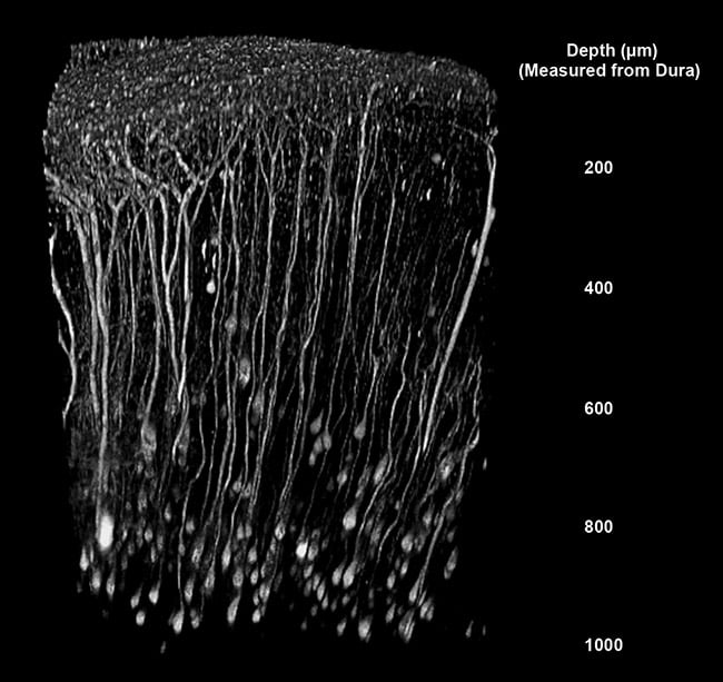
Figure 4. A two-photon image of mouse brain, showing details at 1-mm depths. Courtesy of Mario
Merlini/Akassoglou Laboratory/Gladstone Institute of Neurological Disease.
Technologies converge
As stated, disruptions in blood flow can be measured by monitoring the degrees of oxygenation in hemoglobin. When cells are healthy, they use oxygen at a well-defined rate. When there is a disruption in blood flow, this rate deviates from homeostasis. These differences can be correlated to both stroke occurrence and the repair mechanism. Using multiphoton microscopy, the blood flow can be directly monitored in real time using fluorescent markers9.
In these experiments, a dye is injected into the specimen and the flow velocity is measured directly with submicron spatial resolution. Prior to performing any in vivo experiments, this monitoring is carefully calibrated to ensure that the changes in observed fluorescence arise from blood flow. With this method, disruptions in blood flow will be visible. Combining these multiphoton microscopy and laser technologies provides quantitative measurements of structure and function, with the main limitation being penetration depth. However, both laser and microscope manufacturers continue to push these technologies to longer wavelengths and higher energies to overcome these challenges.
Multiphoton microscopy tells us whether a stroke has occurred, where it occurred, and its severity. Advancements in femtosecond laser and microscope technology allow access to a wide range of fluorescent markers, and the information yields answers to many key questions simultaneously. Most importantly, several researchers have studied the relationship between blood flow and neural activity10 and to what extent the disruption of blood flow affects brain function. Through the use of continuous-wave, picosecond, and femtosecond lasers, researchers are designing experiments to monitor stroke in a clinical setting and in the laboratory.
Together with MRI and CT structural analyses, NIR spectroscopy provides valuable information regarding stroke occurrence, severity, and prognosis, and it plays a key role in deepening our understanding of stroke. How long do nerve cells continue to function following disruption? How does the brain recover, as it often does in many patients? Modern NIR spectroscopy has laid the foundation to answer these key questions.
Meet the author
Ian Read, Ph.D., is senior manager of product marketing for Spectra-Physics scientific lasers at MKS Instruments. During his 20 years with Spectra-Physics, Read has held several positions in sales and marketing. In his current role, he manages a group that markets lasers used for scientific research and bioimaging. Read received his doctorate in physical chemistry from the University of Pittsburgh in 2009 under the direction of David H. Waldeck, where he studied condensed-phase time-resolved (femtosecond and picosecond) spectroscopy of molecular complexes; email: [email protected].
References
1. Centers for Disease Control and Prevention. National Center for Health Statistics, CDC WONDER. Multiple causes of death 1999-2019, https://wonder.cdc.gov/mcd-icd10.html.
2. American Heart Association Council on Epidemiology and Prevention Statistics Committee and Stroke Statistics Subcommittee (2021). Heart disease and stroke statistics — 2021 update. Circ, Vol. 143, Issue 8, pp. e254-e743.
3. H.T. Whelan et al. (2020). Functional near-infrared spectroscopy and its clinical application in the field of neuroscience: advances and future directions. Front Neurosci, Vol. 14, p. 724.
4. H.W. Shytz (2015). Near infrared spectroscopy: investigations in neurovascular diseases. Dan Med J, Vol. 62, p. 12.
5. G. Giacalone et al. (2019). Time-domain near-infrared spectroscopy in acute ischemic stroke patients. Neurophotonics, Vol. 6, p. 015003.
6. A. Kassner and Z. Merali (2015). Assessment of blood-brain barrier disruption in stroke. Stroke, Vol. 46, Issue 11, p. 10.
7. A. Sigler and T. Murphy (2010). In vivo 2-photon imaging of fine structure in the rodent brain: before, during and after stroke. Stroke, Vol. 41, Issue 10, Suppl. 1170.
8. A.Y. Shiy et al. (2012). Two-photon microscopy as a tool to study blood flow and neurovascular coupling in the rodent brain. J Cereb Blood Flow Metab, Vol. 32, No. 7, pp. 1277-1309.
9. X. Fu et al. (2020). In vivo single-molecule detection of nanoparticles for multiphoton fluorescence correlation spectroscopy to quantify cerebral blood flow. Nano Lett, Vol. 20, No. 8, pp. 6135-6141.
10. J. Wu et al. (2020). Kilohertz two-photon fluorescence microscopy imaging of neural activity in vivo. Nat Methods, Vol. 17, pp. 287-290.