New technology enables accurate visualization of internal tissues beyond the reach of other equipment.
HAMID PAHLEVANINEZHAD, HARVARD MEDICAL SCHOOL
The emergence of innovative imaging techniques has enabled several medical applications in detection, diagnosis, and monitoring of diseases. However, there still exists a gap in the imaging of internal organs where tissues are not readily accessible. The inability to accurately visualize the structure and function of tissues in vivo impedes accurate screening, diagnosis, and guiding/monitoring of diseases in internal organs. Clinical studies for understanding pathogenesis are mostly histological; they contain very limited spatial and temporal information and do not provide a view of broad morphological changes caused by diseases. An imaging system capable of accurately visualizing the structure and function of tissues in vivo is crucial in overcoming these clinical hurdles.
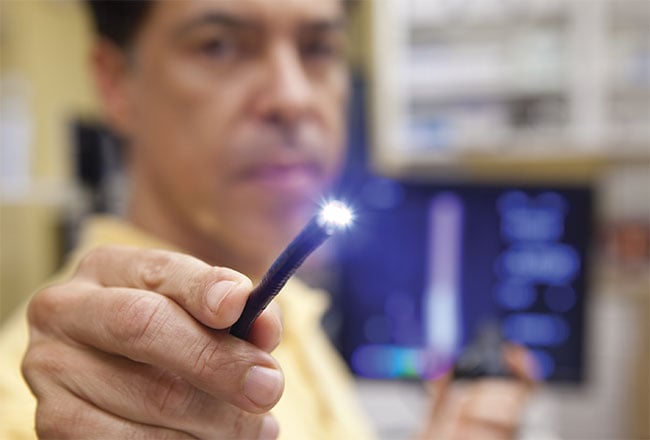
Improvements in endoscopic techniques could provide a complete picture of internal organs and tissues in medicine.
Accurate diagnosis requires visualization of tissue structures at microscopic levels, and optical imaging is a suitable technique to provide sufficient resolution. However, the limitations in the designs of current endoscopic imaging catheters deny the full potential of optical imaging in current endoscopes. The greatest challenge in endoscopic optical imaging is the miniaturization and integration of optical imaging technologies into endoscopic equipment. High-resolution optical imaging through fine focusing of light requires a sophisticated optical design similar to that of a microscope objective, which is difficult to integrate into a miniature endoscope. Even if high-quality focusing is achieved, high-resolution imaging can be maintained only in a very limited depth range because of the trade-off between lateral resolution and depth of focus. Current endoscopic imaging techniques bear significant aberrations — including spherical, astigmatism, and comatic aberrations — that impose severe limits on the resolving power of such endoscopes. Pathological features are often very subtle and easily overlooked, and the limitations in the imaging resolution and depth range of current imaging catheters impede acquisition of conclusive diagnostic information from tissue. For example, adenocarcinoma, the predominant lung cancer cell type, is associated with fine irregular glandular patterns that are difficult to identify in vivo and in real time because of the limits in the resolving power of current techniques1.
Nano-optic endoscopes
Researchers at Harvard University and Massachusetts General Hospital have developed a new class of endoscopic imaging catheters — termed “nano-optic endoscopes2” — that use metalenses3,4 to overcome the limitations of current
imaging systems. Metalenses are ultra-thin optical components composed of nanostructures whose geometry and distributions can be tailored to control light at subwavelength levels. A metalens can impart a precise and predefined phase into the incoming light. This feature allows the nano-optic endoscope to achieve diffraction-limited imaging by negating
aberrations. The nanostructures of a metalens can also be engineered to render different chromatic dispersion properties.
The ability to engineer the chromatic dispersion in the context of spectral interferometry is used to achieve nano-optic endoscopes with greatly extended depth of focus, enabling the maintenance of high-resolution imaging in a large depth range into the tissue. Notably, this outcome is infeasible using conventional techniques where the chromatic dispersion is immutable and at the mercy of the material properties of focusing elements (chromatic dispersion of glass).
Figure 1 shows a nano-optic endoscope for optical coherence tomography. In this example, a prism redirects light to the side for a side-viewing catheter suitable for imaging luminal organs. A metalens focuses light into a diffraction-limited spot outside a sheath. Volumetric images are captured as the optical assembly is spinning and translating within the stationary sheath. Diffraction-limited focusing is achieved when the metalens imparts a phase profile that nulls optical path length differences of ray trajectories from the fiber to the focal point through the metalens. Crucially, the metalens can be designed to impart the exact phase to remove aberrations and achieve diffraction-limited focusing, a feature unavailable to other techniques. The phase profile is realized using amorphous silicon nanopillars with the same height and varying radii modeled through computational wave analysis in the wavelength range of interest (1.25 to 1.37 nm).
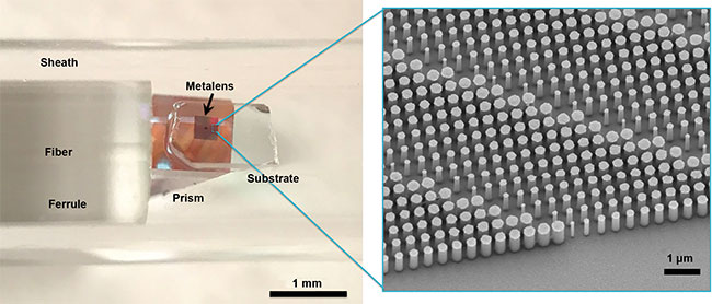
Figure 1. A nano-optic endoscope for optical coherence tomography that uses a length of single-mode fiber housed within a drive cable and ferrule
to deliver and collect light to and from the tissue. Courtesy of Hamid Pahlevaninezhad and Yao-Wei Huang.
The metalens is fabricated using a top-down lithographic approach2 developed by professor Federico Capasso’s group at the Harvard John A. Paulson School of Engineering and Applied Sciences. Briefly, a layer of amorphous silicon will be deposited on a glass substrate. The sample is then spin-coated with a layer of electron beam resist. Nanostructure mask patterns are defined using electron beam lithography followed by deposition of an aluminum layer by thermal evaporation and lift-off. The nanostructures are created using inductively coupled plasma-reactive ion etching.
The optical components at the distal end of the nano-optic endoscope are assembled using a custom-built platform composed of holders for the metalens, fiber, and a prism mounted on separate precision translational and rotational stages to ensure accurate alignment. The components are bonded using optical adhesive during simultaneous monitoring of the output beam to achieve optimal performance.
Figure 2 shows the intensity profiles of the nano-optic endoscope output beam. Focal spot profiles are symmetric and shift axially with wavelength because of the chromatic dispersion of the metalens. This causes spectral interferometry with nano-optic endoscopes to yield imaging with effective axially shifted, overlapping field distributions and, consequently, much larger depth of focus than when an achromatic lens is used in similar circumstances.
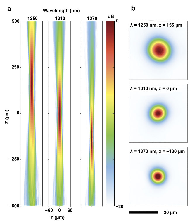
Figure 2. The intensity profiles of the nano-optic endoscope output beam in the tangential (a) and focal (b) planes. Courtesy of Hamid Pahlevaninezhad and Yao-Wei Huang.
An analytic approach is used to model the spectral interferometry with the nano-optic endoscopes2 to assess the effects of chromatic aberrations on imaging depth of focus. The analytic framework includes calculation of the excitation field using a Fresnel-Kirchhoff integral and the insertion of this field into the standard spectral interferometry formalism5 to obtain the lateral resolution at different depth points. The calculated imaging point spread function predicts substantially increased imaging depth of focus (>threefold) for the nano-optic endoscope compared to that when an achromatic lens with the same lateral resolution is used (Figure 3a, b). The increased imaging depth of focus demonstrates yet another remarkable property of the nano-optic endoscope; namely, the flexible chromatic dispersion that can be used to maintain high-resolution imaging in an extended depth range.
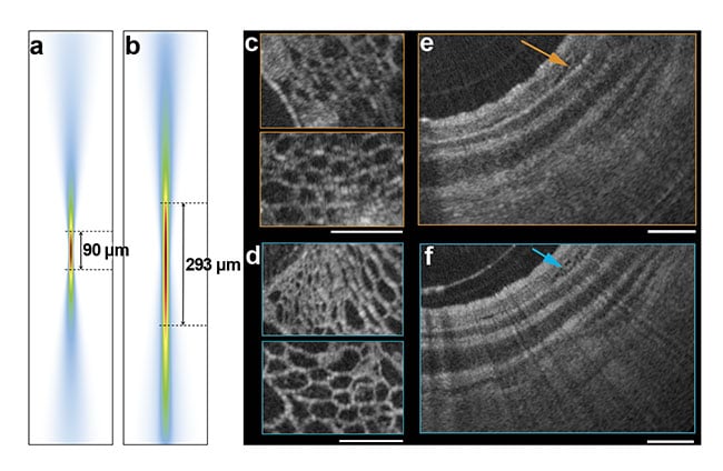
Figure 3. The calculated imaging point spread function predicts substantially increased imaging depth of focus (>threefold) for the nano-optic endoscope (a) compared to that of an achromatic lens with the same lateral resolution (b). A comparison of the imaging quality of the nano-optic endoscope with that of a ball lens catheter (c-f). Courtesy of Hamid Pahlevaninezhad and Yao-Wei Huang.
Figure 3c-f compares the imaging quality of the nano-optic endoscope with that of a ball lens catheter used in commercial endoscopic OCT imaging systems. Imaging was performed on fruit flesh (grape) and swine airways ex vivo. Superior quality of the image of cellular structures in fruit flesh acquired by the nano-optic endoscope is evident when compared to that of the ball lens catheter. In the image of the swine airway, the clear delineation of the layers of the airway wall and the visualization of fine glans (arrow) in the bronchial mucosa further confirm the superior image quality of the nano-optic endoscope in comparison to that of the ball lens catheter.
Design flexibility and versatility of metalenses allow nano-optic endoscopes to achieve greatly improved performance and new functionalities that are unattainable with conventional techniques. These advancements facilitate the translation
of endoscopic optical imaging into wide-spread use in diagnostic applications in medicine by enabling more accuracy in the techniques used.
The ability to control other properties
of endoscope output light, such as polarization states, enables a host of other applications. Several tissues — such as muscle, collagen, and vessel walls — have organized constituents oriented in a specific direction. Polarization-sensitive imaging can contrast these structures from surrounding tissue by detecting their innate birefringence and optic axis. Nano-optic endoscopes designed using polarization-selective metalenses can enable robust endoscopic polarimetry. These capabilities benefit diagnosis and prognosis of several important medical conditions, including asthma6 and fibrosis7.
Meet the author
Hamid Pahlevaninezhad is a research faculty member at Harvard Medical School and Massachusetts General Hospital. His research includes the development of new optical imaging systems with applications in medicine. In particular, Pahlevaninezhad is working on next-generation endoscopic imaging systems with performance beyond the current techniques.
References
1. L.P. Hariri et al. (2013). Toward the guidance of transbronchial biopsy: identifying pulmonary nodules with optical coherence tomography. Chest, Vol. 144, pp. 1261-1268, https://doi.org/10.1378/chest.13-0534.
2. H. Pahlevaninezhad et al. (2018). Nano-
optic endoscope for high-resolution optical
coherence tomography in vivo. Nat Photonics, Vol. 12, pp. 540-547, https://doi.org/10.1038/s41566-018-0224-2.
3. M. Khorasaninejad et al. (2016). Metalenses at visible wavelengths: diffraction-limited focusing and subwavelength resolution imaging. Science, Vol. 352, pp. 1190-1194, https://doi.org/10.1126/science.aaf6644.
4. N. Yu et al. (2011). Light propagation with phase discontinuities: generalized laws of reflection and refraction. Science, Vol. 334, pp. 333-337, https://doi.org/10.1126/science.1210713.
5. A.F. Fercher et al. (1995). Measurement of intraocular distances by backscattering spectral interferometry. Opt Commun,
Vol. 117, pp. 43-48.
6. D.C. Adams et al. (2016). Birefringence microscopy platform for assessing airway smooth muscle structure and function in vivo. Sci Transl Med, Vol. 8, p. 359ra131, https://doi.org/10.1126/scitranslmed.aag1424.
7. L.P. Hariri et al. (2018). Endobronchial optical coherence tomography for low-risk microscopic assessment and diagnosis of idiopathic pulmonary fibrosis in vivo. Am J Respir Crit Care Med, Vol. 197, pp. 949-952, https://doi.org/10.1164/rccm.201707-1446LE.