A superresolution nanoscope could enable 3D imaging of amyloid plaques — a characteristic feature of Alzheimer’s disease — with up to 10 times greater detail than conventional microscopes.
Researchers at Purdue University combined active shaping of point spread functions and adaptive optics to enable robust 3D single-molecule switching nanoscopy (SMSN) imaging within tissues. They used the technology to image through 30-μm-thick brain sections in a mouse model of Alzheimer’s disease. They were able to visualize and reconstruct the morphology and the nanoscale details of amyloid-β filaments.
The nanoscope demonstrated the ability to reconstruct the whole tissue, its cells, and cell constituents at a resolution six to 10×
higher than traditional microscopes.
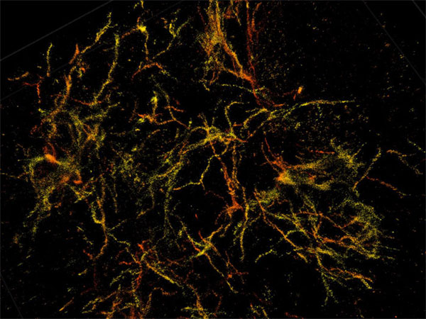
Purdue researchers have taken 3D single-molecule superresolution images of the amyloid plaques associated with Alzheimer's disease in 30-µm-thick sections of the mouse's frontal cortex. Courtesy of Purdue University/Fenil Patel.
The use of adaptive optics compensates for aberrations that can occur when light travels through different parts of a cell or tissue structure at different speeds. To image brain tissue, researchers developed a technique that adjusts the mirrors in response to sample depths. The technique can compensate for aberration and also introduce aberration to maintain the position information carried by a molecule.
The limited resolution in traditional light microscopes and the natural thickness of brain tissue have made it challenging for researchers to clearly observe 3D morphology of amyloid plaques and the plaques’ interactions with other cells.

“Brain tissue is particularly challenging for single-molecule superresolution imaging because it is highly packed with extracellular and intracellular constituents, which distort and scatter light — our source of molecular information," said professor Fang Huang. "You can image deep into the tissue, but the image is blurry.”
Using genetically engineered mice, researchers developed plaques that are characteristic of Alzheimer's disease. Through 3D reconstructions, they observed that the amyloid plaques entangled surrounding tissue via small fibers that branch off waxy deposits.
“We can see now that this is where the damage to the brain occurs," said professor Gary Landreth. "The mouse gives us validation that we can apply this imaging technique to human tissue.”
Researchers have begun using the nanoscope to observe amyloid plaques and how they interact with other cells in human brain samples. The team hopes that the technology can be used to help further understanding of neurological diseases, such as Parkinson’s, MS, and Alzheimer’s.
“While strictly a research tool for the foreseeable future, this technology has allowed us to see how the plaques are assembled and remodeled during the disease process," Landreth said. "It gives insight into the biological causes of the disease, so that we can see if we can stop the formation of these damaging structures in the brain.”
The research was published in Nature Methods (doi:10.1038/s41592-018-0053-8).
Researchers discuss how a new imaging technique could reveal why Alzheimer's disease starts. Courtesy of Purdue University/Erin Easterling.