Stanford University researchers have developed a method to create needle-shaped laser beams of varying lengths and diameters, capable of increasing the quality of images obtained by OCT and other imaging modalities. Adam de la Zerda, research team leader from the Stanford University School of Medicine, said that applied to OCT, the needle-shaped beams extended the depth of focus and improved lateral resolution, signal-to-noise ratio, contrast, and image quality over a long depth range.
In the past, implementing a specific needle-shaped beam has been difficult due to the lack of an established flexible generation method, de la Zerda said. The researchers used the method described in the recent work to create a variety of beams at varying specifications, such as one at an extremely long depth of field or one that is smaller than the diffraction limit of light.
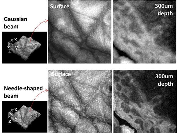
The researchers demonstrated their approach for generating needle-shaped beams by using a needle-shaped beam, 300 μm in length and 3 μm in diameter, to perform OCT imaging of human skin. Their images showed much higher resolution (bottom) than OCT images using a traditional Gaussian-shaped beam. Courtesy of Jingjing Zhao/Stanford University School of Medicine.
OCT features an axial resolution that is constant along its imaging depth. However, its axial resolution — which is determined by the light source — has a very small depth of focus. To address this issue, OCT instruments are often made so that the focus can be moved along the depth to capture clear images of an entire region of interest. However, this dynamic focusing can make imaging slower and doesn’t work well for applications where the sample isn’t static. Additionally, OCT imaging typically uses an objective lens that generates one focal point with a single, short depth of focus.
To increase the depth of focus, the researchers used a phase mask, a diffractive optic element that uses microstructures to create various light patterns resulting in numerous focal points along the axial direction. The phase mask was designed with groups of randomly distributed and specially patterned pixels to create a new focus different from the original one. The entire phase mask generated densely spaced foci in the axial direction, forming a needle-shaped beam with a long depth of focus.
“Flexibility is the primary advantage of this new approach,” said first author Jingjing Zhao. “Both the beam length and its diameter can be flexibly and accurately changed by modifying the locations of the foci and the phase difference between every two adjacent foci.”

The flexibility is made possible by a computational model the researchers created to reveal the relationship between the beam properties and the design parameters of the multiple foci in a precise, quantitative way. They also developed a high-performance fabrication procedure to make diffractive optical elements based on the model’s calculations.
To test their model, the researchers created beam shapes suitable for imaging several different types of samples. For example, to image individual cells within an entire layer of human epidermis, they created a needle-shaped beam with a diameter smaller than 2 μm (cellular resolution) and a length of at least 80 μm (epidermis thickness). They also captured high-resolution dynamic images of a beating heart in a living drosophila larva, an important model organism for studying heart disease. This required a beam that was 700 μm long and 8 μm in diameter to visualize organ structure over a long depth range.
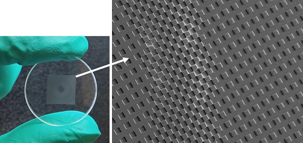
To increase depth of focus, the researchers created numerous focal points with a diffractive optical element known as a phase mask that contains microstructures used to create various light patterns. Courtesy of Jingjing Zhao/Stanford University School of Medicine.
“The rapid high-resolution imaging ability of needle-shaped beams can also get rid of adverse effects that occur due to human movements during image acquisition,” Zhao said. “This can help to pinpoint melanoma and other skin problems using OCT.”
The researchers are working to improve the approach by replacing the diffractive optical element and objective currently used to make a needle-shaped beam with a single flat metalens based on their model. This metalens could be placed on the skull of a mouse to observe the neuron dynamics inside the mouse brain in real time, for example.
The work could also find applications beyond improving OCT, the researchers said. Needle-shaped beams can be used to improve the resolution of microscopy systems, including particle manipulation with optical tweezers, materials processing, photolithography, and photoacoustic tomography, Zhao said. According to Zhao, the model can also be applied to electromagnetic waves for terahertz imaging.
The research was published in Optica (www.doi.org/10.1364/OPTICA.456894).