As systems become more standardized and reliable, OCT expands beyond ophthalmology into other disciplines.
PIERRE FEREYRE AND ANTOINE ADAM, TELEDYNE E2V
Optical coherence tomography (OCT) is a noninvasive imaging technique that is used to deliver high-resolution images in two or three dimensions. It relies on the wave-like properties of light to produce a low-
coherence interference spectrum. In recent decades, this approach has proved to be very beneficial in industry and in medicine. In 1992, a startup company called Advanced Ophthalmic Devices, which grew out of a collaboration at MIT, first used an OCT system as a medical imaging technique for ophthalmology. Since that time, the technology has expanded to enable better understanding and detection of ophthalmic conditions and diseases, including glaucoma, cataracts, diabetic retinopathy, pigment cell detachment, and retinal detachment (Figure 1).
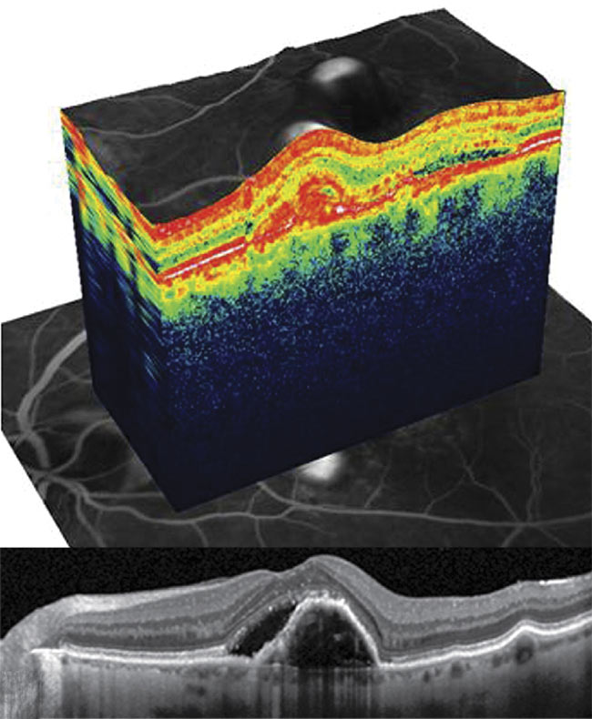
Figure 1. A cross-sectional slice and 3D view of a pigment epithelial detachment resulting from choroidal neovascularization with neovascular age-related macular degeneration. Courtesy of the Eye Clinic in Lodz, Poland/Heidelberg Engineering.
For the majority of eye diseases, early diagnosis is the key to preserving a patient’s long-term vision. Therefore, an efficient OCT system that can be used for a large part of the population has real advantages for patients and professionals, reducing insurance costs and helping to preserve the sight of millions of people. Early identification of eye disorders can be performed by using a so-called primary care OCT system, which operates with the same imaging standards as a mainstream OCT machine, but it uses standard components and focuses on detection prior to diagnosis.
After a long maturation of the technology, OCT has become more accessible because reliable systems can be built at a relatively low price using off-the-shelf components. This has resulted in the emergence of a number of OCT applications, as well as other health segments such as eye disease therapy and prevention. So, what role will OCT play in the future?
More than 100 companies offer OCT systems to address medical and industrial applications, as well as to specifically promote eye health. The adoption of this modern technique is not limited to the field of human health. For example, veterinarians used Bioptigen’s Envisu OCT system to perform surgery on the cataract of C’sar, an elephant residing in a North Carolina zoo.
A key component of the OCT system is the image-capture element. For many years, companies such as Teledyne e2v have been producing line-scan cameras, which are useful in this application for their sensitivity (quantum efficiency) and scanning speed. The camera line rate is a key to reducing the duration of an exam, which provides more comfort to patients while avoiding the appearance of artifacts in the resulting images.
Fourier domain OCT
The signal captured by the camera is not directly usable as in conventional imaging. The optical system produces a spectrogram resulting from the different optical interferences of the light beam in the layers of the eye. The reconstruction of the final three-dimensional image passes through a mathematical processing called Fourier transform to provide depth information. This approach is referred to as Fourier domain OCT (FD-OCT). There are two main families of systems using the same principle: spectral domain OCT (SD-OCT) and swept-source OCT (SS-OCT), also called optical frequency domain imaging. They differ essentially in the method of construction of the spectrogram and in the optomechanical components employed. SD-OCT uses broadband and low-coherence light sources, while SS-OCT uses coherent, narrow-band light.
SS-OCT has disadvantages, one of which is its higher cost when compared to SD-OCT. SS-OCT uses a longer wavelength of light, achieving deeper penetration. It requires narrow linewidth, high speed, and frequency-swept lasers, which can be pricey. Other disadvantages of SS-OCT compared with SD-OCT include lower axial resolution, a worse signal-to-noise ratio, and worse motion artifacts.
As a consequence, the justification of using SS-OCT is highly dependent on the clinical requirement for the diagnosis of particular visual diseases. SS-OCT enables deeper penetration in tissues because it works at a slightly higher wavelength than SD-OCT, and it is efficient at penetrating through cataracts or hemorrhages.
SS-OCT and SD-OCT systems have in common an approach based on the conjugation of a reference signal (reference arm) and the light reflected by the sample (sample arm) at the output of a Michelson interferometer. The choice of the light source is determined by the expected performance, such as axial resolution:
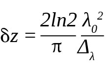
with the central wavelength typically 850 nm and the light spectral bandwidth typically 50 nm.
Each interface of the various layers of the sample backscatters the light in the sample branch, dephasing with the reference branch. The combination of the two signals results in an interferogram pattern characterizing the sample — the thickness and nature of the different layers of organic tissue. One of the key subelements of the SD-OCT system is the combination of the diffraction grating and digital camera components that constitute the spectrometer. This is critical to the accuracy of the system, which is directly dependent on producing a spectrogram with a high signal-to-noise ratio. For example, the implementation of the latest CMOS technology used in the image sensor provides much better performance in terms of dynamic range and spectral sensitivity, which enhances the analysis capabilities. In addition, the spatial resolution of the sensor tends to increase either for a better accuracy or for a greater depth of analysis. The number of pixels sampling the spectrum determines the maximum depth of the image, as follows:
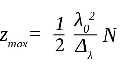
with N as the given number of pixels of the linear image sensor.
Typically, a CMOS image sensor with a resolution of 2048 pixels achieves an imaging depth of 1.5 mm, which is quite suitable for the cross-sectional view of the human retina.
The Fourier transform of the interferogram produces a one-dimensional depth map (A-scan). A displacement of the light beam on the sample is realized (thanks to a two-axis mirror) and produces a full three-dimensional cartography image by combining the A-scan. The observation of the image of the sample is then possible in three-dimensional (point cloud) form or a two-dimensional (cross section) view. The application requires a scan in a sufficiently short time to sustain the patient’s comfort and to obtain artifact-free images. Typically, each A-scan is performed in a time interval of less than 1 ms to allow full examination in less than 1 s.
One of the main benefits of SD-OCT camera technology is that it can be specifically tuned to provide users with the most accurate images — for example, of the retina and cornea — mapping tissue structures, measuring thickness, and visualizing blood flow dynamics for diagnostic purposes. Other advantages include a low power consumption system (reduced by 60% compared to previous designs), operation at constant temperature for reproducible results, and minimal maintenance.
Neurogenerative diseases
Ophthalmology has been the sector in which OCT has matured and consolidated during the past decade. But, in the future, OCT will likely be used for many scientific and medical applications, including cardiology, oncology, and dermatology. A range of companies — from startups to large, established companies — are offering technologies and equipment for the examination of brain cells, where it may be possible to detect the early signs of diseases such as Alzheimer’s or Parkinson’s. These new fields of application are very promising for the treatment of common neurodegenerative diseases that affect the lives of millions of people around the world.
An interesting illustration of an emerging OCT application can be found in a project undertaken by Duke University. Beth Doyle, head of the Conservation Services Department for Duke University Libraries; Michael Toth, project manager at Duke University Health System; and Sina Farsiu, assistant professor of biomedicine and ophthalmology at Duke, used an OCT imaging scanner to analyze a strip of second-century BCE Egyptian mummy cartonnage to see whether there was any hidden text on the papyrus (Figures 2 and 3).

Figure 2. A collaboration at Duke University used an OCT imaging scanner to analyze a strip of second-century BCE Egyptian mummy cartonnage. Courtesy of University College London.
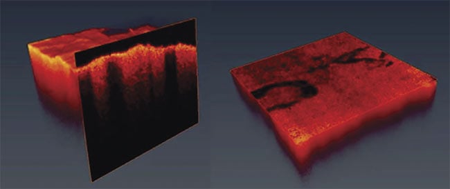
Figure 3. OCT was used to reveal Greek text hidden inside the papyrus of an Egyptian mummy.
Courtesy of University College London.
If OCT equipment can be useful in ophthalmology, it follows that the non-
destructive approach is also of practical use to archaeologists — for example, to see inside fragile papyrus dating from antiquity. Toth examined the fragments using the OCT imager and found that Greek text was written inside. The characters were undetectable by the human eye and revealed that Greek was the language of the government during the age of the more than 2000-year-old papyrus.
OCT is a continuously evolving technique. The latest progress in this field involves combining artificial intelligence (AI) with OCT to bring significant benefits to eye disease diagnosis. In 2016, the AI company DeepMind announced a partnership with Moorfields Eye Hospital NHS Foundation Trust in London to develop an AI technology that could quickly analyze complex OCT scans (a time-consuming task for human experts), recommend a referral, and then prioritize patients — as accurately as, or better than, world-leading expert doctors.
The first results comparing machine versus human show promise. It is reported that when a practitioner uses the AI system, the diagnostic error rate is equivalent to the rate of a retina specialist with 21 years of experience. In the case of an entry-level optometrist with two to three years of experience, the machine is four to five times more efficient. This technological advancement is a very promising step forward, as the productivity gains will allow a wider deployment of diagnostic tests and the identification of patients requiring urgent treatment, preventing loss of eyesight for many.
Therefore, the adoption of the OCT technique on a large scale — with the objective of early prevention of eye and other diseases — represents a societal challenge with worldwide scope and could be a major development in maintaining the health of populations.
Meet the authors
Pierre Fereyre is a sensor design specialist for Teledyne e2v in Grenoble, France; email: [email protected].
Antoine Adam is a marketing specialist for Teledyne e2v in Grenoble, France; email: [email protected].