Close

Search

Search Menu
Photonics Media Photonics Marketplace Photonics Spectra BioPhotonics Vision Spectra Photonics Showcase Photonics ProdSpec Photonics Handbook

Olympus Announces Light Microscopy Image Awards

Facebook Twitter LinkedIn Email
Olympus has announced the winners of its first Global Image of the Year Life Science Microscopy Award.

The winning image, captured by Ainara Pintor of Spain using a super resolution confocal microscope system The image shows the immunostaining of Thy1-EGFP mouse brain slice with two fluorophores. In green, the excitatory hippocampal neurons, which express Green Fluorescent Protein under Thy1 promoter. In red, Fat mass and obesity-associated (FTO) protein revealed with Alexa Fluor 594 antibody. In blue, cell nuclei labelled with DAPI. Courtesy of Olympus.
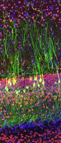
The winning image, captured by Ainara Pintor of Spain using a superresolution confocal microscope system. The image shows the immunostaining of a Thy1-EGFP mouse brain slice with two fluorophores. In green, the excitatory hippocampal neurons, which express green fluorescent protein under Thy1 promoter. In red, fat mass and obesity-associated (FTO) protein revealed with Alexa Fluor 594 antibody. In blue, cell nuclei labeled with DAPI. Courtesy of Olympus.

Ainara Pintor of Spain earned first place for her vibrant image of an immunostained mouse brain slice with two fluorophores, titled “Neurogarden.”

“There are over 70 million neurons in a mouse brain,” Pintor said. “This is an example of what we can observe in the hippocampus of a single brain slice, in this case, taken from Thy1 transgenic mice.”

For the grand prize, she will choose either an Olympus CX43 microscope with a DP27 digital camera, or Olympus’ X Line objectives.

In addition to the global award, regional prizes were awarded to Howard Vindin of Australia, Tagide deCarvalho of the United States, and Alan Prescott of the United Kingdom. Each regional winner will receive an Olympus OM-D E-M5 Mark II digital camera. Honorable mentions went to Ming-Der Lin, Nat Prunet, Justin Zoll, Tong Zhang, Daniela Malide, Hamed Rajabi, Rudolf Buechi, Martin Hailstone, and Nathan Renfro.

Images were evaluated based on artistic and visual aspects, scientific impact, and microscope proficiency.

To learn more about the images and the microscope techniques used to capture them, visit www.olympus-lifescience.com/ioty.


Photonics Spectra
Jun 2020
GLOSSARY
fluorescence
The emission of light or other electromagnetic radiation of longer wavelengths by a substance as a result of the absorption of some other radiation of shorter wavelengths, provided the emission continues only as long as the stimulus producing it is maintained. In other words, fluorescence is the luminescence that persists for less than about 10-8 s after excitation.
fluorescence microscopy
Observation of samples using excitation produced fluorescence. A sample is placed within the excitation laser and the plane of observation is scanned. Emitted photons from the sample are filtered by a long pass dichroic optic and are detected and recorded for digital image reproduction.
BusinessBiophotonicsfluorescencefluorescence microscopyconfocal microscopyOlympusawardsMicroscopyRapidScanlight speed

back to top
Facebook Twitter Instagram LinkedIn YouTube RSS
©2023 Photonics Media, 100 West St., Pittsfield, MA, 01201 USA, [email protected]

Photonics Media, Laurin Publishing
x Subscribe to Photonics Spectra magazine - FREE!
We use cookies to improve user experience and analyze our website traffic as stated in our Privacy Policy. By using this website, you agree to the use of cookies unless you have disabled them.