A rising number of light-sensitive proteins and optogenetics are enabling precise imaging of brain cells, as well as the potential to adapt functioning in neural networks.
The brain is one of the most complex systems in the known universe, a living computer that controls not only basic functions of the body but ponders and directs responses to what’s going on in its surroundings. But when it comes to understanding how the brain works — and, in some cases, how to heal what is ailing it — practitioners of optogenetics know that the answer can lie in natural optical phenomena that have existed since before modern life even began.
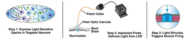
In optogenetics, researchers and clinicians introduce a series of light-sensitive proteins that can effectively transmit DNA to various types of neural cells via a natural pumping mechanism within the molecules. The light-sensitive properties can thus be transferred.
Orange light bathing a brain and silencing cone opsins. Courtesy of Ed Boyden/McGovern Institute.
These transmembrane proteins, called rhodopsins, are common in many forms of life, such as algae, and they also help to initiate photosynthesis in other microbes. Similar molecules are at work in the eyes of humans and other animals, according to Ed Boyden, professor at MIT’s McGovern Institute for Brain Research in the Department of Brain and Cognitive Sciences, and in the Department of Biological Engineering. When introduced through a stereotaxic procedure and excited by light, the interaction of rhodopsins with nearby neurons can enable control of specific processes in the brain. And when the proteins are used to transform neighboring cells, they can at times reinvigorate or inhibit brain functions.
Science makes history
Opsins were first activated in the brains of specimens in the laboratory in 2004, thanks to the work of Boyden, professor Karl Deisseroth at Stanford University, and researchers working simultaneously at other institutions. A surge of molecular engineering and genome prospecting, partially driven by the investigation of various species of algae, led to the discovery and creation of opsins that were functional for particular purposes. Synthetic opsins have since been developed that result in controlling specific biochemical events in freely moving animals1.
Now the inventory of opsins has multiplied from the seven originally identified to many times that. Boyden differentiated between channelrhodopsins, halorhodopsins, and archaerhodopsins, which all have unique purposes for the optogeneticist. Generally, channelrhodopsins incite electrical activity of neurons, while the latter two reduce that activity.
The work of Deisseroth and colleagues has been groundbreaking, but it has not happened in a vacuum, according to Valentina Emiliani, interdisciplinary research director at the Vision Institute in Paris. In a lecture at Neuroscience 2019 in Chicago, she said innovations in microscopy, imaging, and holographic light shaping in recent decades have opened the door to stimulation on the single-cell level, as well as precise mapping of brain functioning.
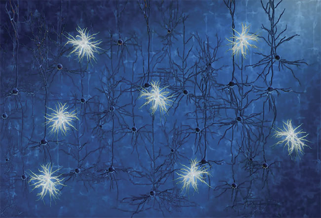
When blue light hits a neural network in which one kind of neuron is light sensitive, only that neuron
is activated. Courtesy of Ed Boyden/McGovern Institute.
“I would definitely say that two-photon excitation combined with light shaping and opsin engineering have enabled us to enter a new phase of optogenetics where it is possible to manipulate neuronal circuits at cellular resolution (we called this ‘circuit optogenetics’) not possible by using wide-field single-photon illumination,” Emiliani said.
And indeed, in a research paper published last year, she and colleagues reported that by using a high-powered laser and low intensity, they could focus on specific areas of mouse brains. With the benefits of two-photon holographic excitation, electrophysiological recordings, and calcium imaging, they were able to control — with millisecond temporal resolution — neuronal activity via three opsins2.
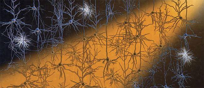
Light-activated opsins can excite or reduce activity in the primate brain to help identify various brain
functions. Courtesy of Deniz Dalkara/Vision Institute.
“The interest of this still-growing number of new opsins is in the fact that they have different absorption spectra so that they can be combined with calcium indicators to perform all-optical neuronal manipulation,” Emiliani said. “This is important to avoid cross talk — that is, the artifactual excitation of the opsins by the imaging laser. They also have fast kinetics and they can be soma [nucleus] restricted and so enable true single-cell resolution.”
In some instances, according to Boyden, various opsins have been used in the brains of laboratory animals to redirect dopamine neurons in reward centers to modify behavior.
“There is no perfect treatment, but when we use pharmaceuticals to treat a condition in the brain, we’re basically bathing the brain in a chemical, which is why there can be side effects,” he said. “Electrical stimulation can be effective, but electricity goes in all directions. Optogenetics, using optical fibers connected to lasers, can activate specific neurons in the brains of laboratory animals. Then these data can inspire better pharmaceuticals and electrical stimulation procedures for human treatment.”
Boyden said the introduction of these opsins can be unnatural, too: “It is not normal to have a molecule from algae in the brain.” Nevertheless, no repercussions of this process have been identified so far.
Tools of the trade
As the science of optogenetics has matured, the toolkit for using it has expanded, with new and varied equipment now on the production line. Much of this development has been promoted by the government-sponsored BRAIN Initiative
(Brain Research Through Advancing Innovative Neurotechnologies), under the purview of the National Institutes of Health, with a wide array of companies and research institutions involved.
Researchers now have the benefit of plug-and-play equipment to make use of various aspects of the process of optogenetics both in vivo and in vitro. Thorlabs Inc., for example, offers a whole line of components — most often used in research labs — including LEDs, patch cables, rotary joints, interconnects, cannulae, and stereotaxic tools that can be purchased separately or as a complete system. The resolution created with this equipment can keep up with real-time neural activity and enable various wavelengths to energize different neurons at the same time.
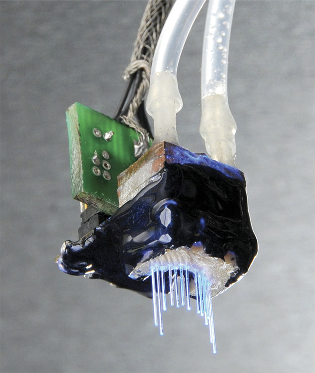
An array of optical fibers, each connecting to an LED, enables light to be delivered to multiple deep targets in the brain. Courtesy of Jake Bernstein and Ed Boyden.
“Each wavelength will have a different light source associated with it, with LED sources being the most common variety,” said Patrick Hawkins, a design engineer at Thorlabs. “There are ways to combine sources — for example, a fiber optic coupler — where different branches can be connected to different light sources but then combined together to a common output.”
Equipment from various manufacturers could be cross-compatible, depending on specifications. The fiber core diameter and numerical aperture need to match, he said.
When a system is designed and used correctly, it can have a powerful impact on the activity of specific neural cells. In particular, Hawkins pointed to a channel rhodopsin (ChR2) at 470 nm, which has gained popularity in the practice of optogenetics.
An optogenetics experiment on a white mouse brain. Courtesy of Thorlabs.
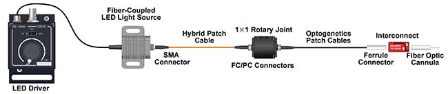
The equipment involved in an optogenetics setup. Courtesy of Thorlabs.
“A popular application would be to combine two wavelengths to alternatively excite and inhibit a behavior or to study two totally different behaviors,” he said. “You can do this with two fiber optics implanted simultaneously with a common configuration being bilateral stimulation; 590 nm is a popular wavelength to add to a setup, other than 473 nm. The absorption spectra of these opsins have pretty long tails associated with them, so choosing two very different wavelengths like this would be much more effective than using 473 and 490 nm simultaneously, for example.”
Technique evolves
As it turns out, optogenetics has found itself bound to other forms of science in terms of both equipment and research. In some cases, this confluence has resulted in more potent results than any one strategy could produce on its own.
Another team at Stanford, led by Guosong Hong, for example, recently devised a method of using both sound and light to penetrate deep into the brain. The challenge, they said, was that — while optogenetics can be an effective tool — light cannot penetrate deeply, so optical implants must be installed in a subject’s brain, often requiring a craniotomy. The need to perform such an invasive procedure is lessened, however, when combined with ultrasound technology. The researchers were able to inject certain nanoparticles that emit light when activated by ultrasound.
In a recent paper, the team described how they accomplished this result in the laboratory. They reported that so-called mechanoluminescent nanoparticles can
be injected intravenously and act as an
energy relay between superficial blood vessels near the skin and deep vessels in the brain. They used a 400-nm light to charge the nanoparticles as they flowed through the blood vessels. The particles store the energy of the 400-nm excitation during circulation and then release the energy as 470-nm emission as they pass through the ultrasound focus deep in the brain. The brain-penetrant ultrasound therefore “gates” the emission of a 470-nm blue light anywhere and anytime at will in the brain through intact scalp and skull.
The researchers called this process “sono-optogenetics” and reported stimulation in the brains of live mice. The stimulation produced contralateral limb movement that corresponded with focused ultrasound pulses3.
Hong — assistant professor of materials and science engineering and a member of the Wu Tsai Neurosciences Institute at Stanford — devised the approach and supervised the experiments. He said that while the circulating nanoparticles need to be a certain distance from their target neurons, the extensive network of vessels solved the problem. “The falloff distance of 470-nm light, which is produced by the nanoparticles, is about 200 µm in the brain. Since the nanoparticles are delivered by blood circulation, and since the cerebral vasculature is pervasive in the brain with an average distance between the two closest vessels of 100 µm, ultrasound-triggered light emission from circulating nanoparticles can in theory cover every piece of the brain with precise location.”
According to Hong, while the need for reduced-size equipment is a challenge for researchers, invasiveness — one of the main barriers to widespread use of optogenetics — has been decreased to the point where the technology could be introduced into clinical trials in the very near future.
The best may be yet to come, thanks to a convergence of scientific innovation, according to Boyden. With emerging trends such as expansion microscopy and voltage imaging, optogenetics practitioners may be on the cutting edge of a whole new era of the technique. The BRAIN Initiative is nearing completion of its research phase and is moving into implementation. One of the many tracks being pursued is the use of high-definition brain imaging and analyses of the brain’s synapses — and how and where the synapses can be adjusted. This offers profound potential for patients suffering from epilepsy, believed by some to be caused by overactive portions of the brain, and a variety of other brain disorders and injuries.
References
1. F. Zhang et al. (Dec. 23, 2011). The microbial opsin family of optogenetic tools. Cell, Vol. 147, Issue 7, pp. 1446-1457.
2. I. Chen et al. (May 1, 2019). In vivo submillisecond two-photon optogenetics with temporally focused patterned light. J Neurosci, Vol. 39, No. 18, pp. 3484-3497.
3. X. Wu et al. (Dec. 26, 2019). Sono-opto-genetics facilitated by a circulation-delivered rechargeable light source for minimally invasive optogenetics. Proc Nal Acad Sci, Vol. 116, No. 52, pp. 26332-26342.
Clues Found in Primate Research
Optogenetics has been used to create learned behaviors in mice for several years, according to professor José-Alain Sahel at the University of Pittsburgh School of Medicine. The technique has shown the ability to rehabilitate animals that have had parts of their brains damaged. The implications, and the adaptability of opsins, are enormous for the treatment of conditions centering on the brain, with symptoms affecting motor functioning, speech, and vision.
Sahel, who teaches in the Department of Ophthalmology and spoke at a forum on vision restoration at Neuroscience 2019 in Chicago, has advocated strongly for the introduction of these techniques on nonhuman primates (NHPs) to more clearly mimic the effects such experimentation would have on humans. In a recent paper1, Sahel and colleagues pointed out that only primates have a macula (the functional center of the retina) and therefore can perform relatively complex activities to measure awareness and perception. The researchers illustrated the introduction of adeno-associated viruses — and the intraocular pressure that may cause macular edema and glaucoma, among other conditions — as well as corresponding therapies, into primates.
He said that currently half a dozen studies in optogenetics depend on NHP research.
The researchers wrote, “The genus Macaca, as the most widespread NHP, diverged from its ape and human ancestors 25 million years ago. The genus diversified approximately 5 million years ago and includes over 20 species that share >90% DNA sequence similarity and highly conserved protein sequences with humans. Moreover, humans and macaques share susceptibility genes for age-related macular degeneration (AMD) and genotype-phenotype correlations for rare forms of inherited retinal diseases like achromatopsia and retinitis pigmentosa.”
The organization of photoreceptor cells in the macula is very similar between humans and NHPs, they said, with densely packed cones in the fovea (a depression in the inner retina) of up to 160,000 to 200,000 cones per sq mm.“ In vitro preparations allowed us to demonstrate functional expression of genes such as microbial opsins in cone photoreceptors and retinal ganglion cells in the development of optogenetic therapy for restoring vision,” the researchers wrote.
To lessen the impact on the primates during study, Sahel said researchers use organoids (tissue cultures in vitro), mathematical simulation, and a large number of rodent studies to inform their experiments.
Reference
1. S. Picaud et al. (Dec. 26, 2019). The primate model for understanding and restoring vision. Proc Nal Acad Sci, Vol. 116, No. 52, pp. 26280-26287.