Robust physical properties and enabling capabilities make optical fiber-based sensors increasingly attractive for biomedical applications ranging from light guiding and imaging to complex robotic surgery.
Alexis Méndez, MCH Engineering LLC
The biomedical community recognized the advantages of optical fibers long ago, accepting them even before their adoption for long-haul telecommunications1. Early research on the light-guiding properties of fibers in the late 1920s was aimed at applications in medical imaging. The first clad optical fiber was drawn on December 8, 1956, by Larry Curtis (Figure 1), a graduate student under Basil Hirschowitz at the University of Michigan. The technology was used in a multifiber bundle for a fiber endoscope, which Hirschowitz first tested on himself2.
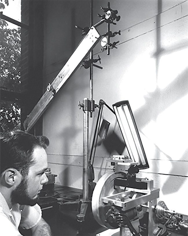
Figure 1. Larry Curtis of the University of Michigan draws the first optical clad fiber by the rod-in-tube method, for use in a medical endoscope. Courtesy of Abraham Katzir.
Optical fibers are attractive for biomedical applications because they are thin, flexible, dielectric (nonconductive), immune to electromagnetic interference, chemically inert, nontoxic, and lightweight. They can also be sterilized using the standard medical sterilization techniques: steam heat, radiation, or dry gas.
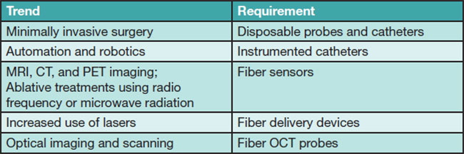
Their adoption in the medical field has expanded widely since the first endoscope, from imaging and analyzing biological functions to enabling rapid advancements in newer medical procedures, such as minimally invasive and robotic surgery (Table 1).
Table 1.
Medical Industry Trends Promoting Increased Use of Optical Fiber
Fiber optic sensors compete with a limited number of conventional electronic devices in biomedical applications. Their primary advantage is the electrical isolation provided by the dielectric optical fibers, which enables safe use without risk of electric shock to a patient. Fiber optic sensors can also be used safely while patients undergo MRI, CT, or any other type of electromagnetic scan, without risk of inducing an electric current or generating heat from the large magnetic fields. Their small size — which enables integrating the sensors along small needles, catheters, and surgical tools — is another key advantage.
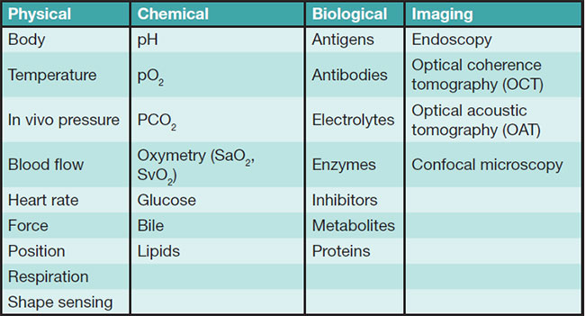
These factors make fiber optic sensors ideally suited for a broad variety of invasive and noninvasive applications in the life sciences, clinical research, medical monitoring, and diagnostics. As shown in Table 2, biomedical optical sensors can be categorized into four main types: physical, chemical, biological, and imaging.
Table 2.
Classifications of Biomedical Optical Fiber Sensors
Optical fibers for imaging
Imaging sensors encompass both endoscope devices for internal observation and imaging, and more advanced techniques — such as optical coherence tomography (OCT), photoacoustic imaging, and others — where internal scans and visualization can be made nonintrusively.
Endoscopic imaging remains the most successful biomedical application for fiber optics.
Optical fibers can also be used to transmit light to tissue regions of interest, either to illuminate the tissue for inspection or to directly cut or ablate it (with a higher-powered laser). Hence, they are used extensively as laser-delivery probes, as well as imaging conduits for diverse illumination applications, either alone or integrated into medical instruments such as otoscopes and laryngoscopes.
Fibers are also used as scanning probes in advanced imaging applications such as OCT and confocal microscopy. And when fused together as plates or tapers, they are used as light-guiding components in digital x-ray devices to guide the light image from a scintillator to an electronic CCD detector array.
Physiological measurements
Physical sensors are used to measure a broad variety of physiological parameters, such as body temperature, blood pressure, respiration, heart rate, blood flow, muscle displacement, and cerebral activity.
Biomedical sensors using external-cavity Fabry-Pérot interferometers, fiber Bragg gratings, and fiber spectrometers based on light absorption and fluorescence are among the fiber optic sensors most commonly developed into commercial products, with temperature and pressure being the most popular parameters. The unique size benefits provided by fiber optic sensors can clearly be appreciated in the pressure and temperature Fabry-Pérot probes shown in Figure 2.
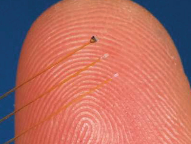
Figure 2. Fiber optic Fabry-Pérot medical pressure and temperature sensors. Courtesy of FISO Technologies.
Some of the earliest fiber optic physical sensors date back to the early 1980s to such pioneering companies as Camino Laboratories, FISO Technologies (now part of Resonetics), and Luxtron (now part of Advanced Energy). In 1984, Camino introduced an intracranial pressure sensor based on an intensity-modulating fiber optic device using a miniature bellows as the transducer. FISO of Canada supplies OEM medical fiber optic pressure and temperature sensors based on external-cavity Fabry-Pérot interferometers interrogated with white light interferometry. OpSens and Neoptix in Canada and RJC Enterprises in the U.S. are other long-standing developers and OEM manufacturers of temperature and pressure fiber optic sensors.
Chemical and biological analysis
Chemical sensors rely on fluorescence, spectroscopic, and indicator techniques to measure and identify the presence of chemical compounds and metabolic variables such as pH, blood oxygen, and glucose. These sensors detect specific chemical species for diagnostic purposes, and they monitor the body’s chemical reactions and activity for diagnostic and therapeutic applications.
Biological sensors tend to be more complex and rely on biologic recognition reactions — such as enzyme-substrate, antigen-antibody, or ligand-receptor — to identify and quantify specific biochemical molecules of interest. One prospective implementation of such sensors is the so-called lab on a fiber3 that combines optical fiber coatings with micro- and nanosize functionalized materials that react to specific physical, chemical, or biological external effects, creating multifunction, multiparameter sensing devices (Figure 3). Light remotely excites the functionalized materials embedded in the fiber’s coating. These materials, in turn, react to specific biological or chemical substances (analytes) and induce an optical signal change proportional to the analyte concentration.
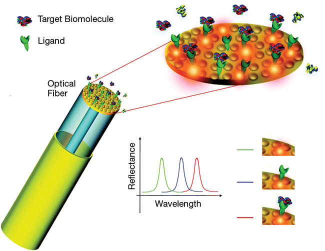
Figure 3. An illustration of the concept of a biomedical lab-on-a-fiber multianalyte sensor. Courtesy of Andrea Cusano.
Biomedical sensors (fiber optic or otherwise) present some unique challenges4. Sensors must be safe, reliable, highly stable, biocompatible, amenable to sterilization and autoclaving, and not prone to biologic rejection. And they must not require calibration, or they must at least be capable of maintaining calibration for prolonged times. Sensor packaging is critical. Small size is highly desirable for these sensors, particularly if they are intended for implanting or indwelling.
Optical fibers for surgical applications
Developments in minimally invasive surgery and remote robotic surgery have widened the market for medical sensors, adding position and force requirements to the sensing regime.
In minimally invasive surgery, surgeons avoid cutting open patients and instead perform small cuts and incisions through which a variety of surgical instruments and catheters are inserted. For remote robotic surgery, the surgical catheters must be fitted with sensors that provide both three-dimensional position information and force (haptic) feedback to the surgeon controlling the robot.
Optical fiber sensors are ideal for this application because they can be easily integrated inside the slender robotic catheters, and, by using multicore fibers inscribed with arrays of fiber Bragg grating strain sensors, it is possible to perform in situ and real-time 3D shape and position sensing. As the fibers bend, twist, and rotate, the fiber Bragg gratings detect the induced strains in the fiber. Specialized algorithms process the data to deliver inverse kinemetrics of spatial position in real time.
One system using such technology is Philips Healthcare’s Fiber Optic RealShape (FORS). Other companies — such as FBGS in Belgium, PhotonFirst in the Netherlands, Intuitive Surgical in the U.S., and Fibercore in the U.K. — are actively working on this type of biomedical fiber optic sensor solution.
Another related innovation is the use of fiber Bragg gratings or Fabry-Pérot elements as force-sensing devices. The functionality of many drug-delivery, therapeutic, and medical device systems relies on a change in force. Force sensors can measure changes in force and relay this information to the clinician and/or patient for adjustments. Fiber optic force sensors, such as Abbott’s TactiCath ablation probe, are available for radio frequency or laser-based ablation devices for treating atrial fibrillation.
Along similar lines, Often Medical in Italy has developed an epidural anesthesia kit based on a fiber Bragg grating force sensor. The system enables real-time monitoring of epidural catheter placement and provides guidance of the needle until it reaches the epidural space, where the incorporated fiber sensor verifies the correct placement of the catheter.
Technical trends and commercial outlook
Optical fibers — and photonics technologies in general — represent a set of very powerful and versatile enabling technologies for medical devices, instrumentation, and techniques for diagnostic, therapeutic, and surgical applications. With a growing population requiring health care and increasingly sophisticated diagnostic tools, clinicians worldwide rely more and more on advanced biomedical instrumentation and sensors as necessary and effective tools for diagnosing, monitoring, treating, and caring for patients.
As costs drop and new sensing techniques are developed, the number and diversity of biomedical fiber optic sensors will increase.
Future trends include the development of increasingly small and thin medical probes and catheters and a proliferation of laser-based treatments and therapies that require fiber optic delivery systems. OCT is also poised to become as common as conventional ultrasound scanning.
Endoscopy will continue to evolve as smaller and more sophisticated devices combine more advanced functions — beyond the standard illumination and visualization — such as direct tissue analysis and laser treatment. Optical imaging techniques will continue to advance, with digital x-rays making noninvasive examination and diagnosis safer and faster due to greater resolution and pinpoint accuracy.
The biomedical sensing market represents a lucrative and growing opportunity for fiber optic sensors, particularly for large volumes of disposable sensing probes. And there is also an unquestionable opportunity for fiber optic sensors to be used as electromagnetic-interference-compatible sensors to monitor patients’ vital signs during the use of MRI (and related techniques) and radio frequency treatments, such as atrial fibrillation ablations. As costs drop and new sensing techniques are developed, the number and diversity of biomedical fiber optic sensors will increase.
Meet the author
Alexis Méndez, Ph.D., is president of MCH Engineering LLC. He received a doctorate in electrical engineering from Brown University and has 30 years of experience in optical fiber technology, sensors, and photonics.
References
1. A. Katzir (1990). Selected Papers on Optical Fibers in Medicine. SPIE Milestone Series, Vol. MS 11. SPIE Press.
2. B. Hirschowitz (1979). A personal history of the fiberscope. Gastroenterology, Vol. 76, No. 4, pp. 864-869.
3. A. Cusano et al., eds. (2015). Lab-on-Fiber Technology. Springer Verlag.
4. É. Pinet and C. Hamel (2007). True challenges of disposable optical fiber sensors for clinical environment. Presented at the Third European Workshop on Optical Fibre Sensors. Proc SPIE, Vol. 6619.