A new optical microscope uses holographic techniques to stimulate multiple cells simultaneously and monitor cell activity after stimulation. Developed by researchers at Kobe University and called SIFOM for three-dimensional (3D) Stimulation and Imaging-based Functional Optical Microscopy, the system consists of two subfunctions: 3D observation of cells and 3D stimulation of cells based on digital holography. SIFOM can precisely stimulate user-defined targeted cells and simultaneously record the volumetric fluorescence distribution in a single acquisition.
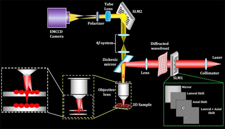
This is a concept drawing of the SIFOM system for cell manipulation technology combining 3D fluorescence observation and 3D stimulation. Courtesy of Kobe University.
SIFOM achieves precise, simultaneous stimulation of fluorescent-labeled cells by using multiple 3D spots generated by digital holograms displayed on a phase-mode spatial light modulator. Single-shot 3D acquisition of the fluorescence distribution is accomplished by digital holographic microscopy. The system uses high-speed scanless photography to obtain information about multiple events occurring in 3D space within a short time frame.
To validate the system, the researchers from Kyoto Institute of Technology and Utsunomiya as well as Kobe University used lung cancer cells and fluorescent beads about 10 μm in size. They recorded a fluorescent hologram in a defocused state from the focal position in the direction of depth and achieved reconstruction of both the cells and the fluorescent beads.
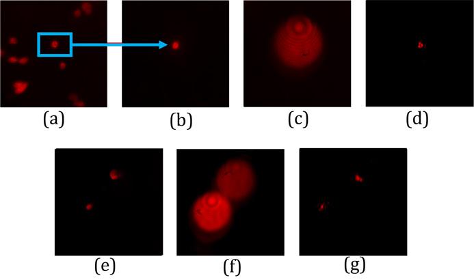
3D recording of lung cancer cells: (a) 2D fluorescent observation, (b) a fluorescent hologram when one cell is selectively extracted, (c) a hologram when moved to a depth of 80 μm, (d) a reconstruction of (c), (e) after extracting two cells and exposing to light stimulation, (f) a hologram when moved to a depth of 80 um, (g) reconstruction of (f). Courtesy of Kobe University.
During the verification experiment, the researchers were able to observe light stimulation for a maximum of five cells at one time. The number of cells that could be stimulated was limited by the amount of light power available for stimulation. In 2D space, the team expects that simultaneous light stimulation will be possible for more than 100 cells. In the future, the team plans to expand the stimulation depth to a few hundred μm using two-photon stimulation.
To avoid damage to living cells, the amount of fluorescence used to observe them must be controlled, the researchers said, adding that high-sensitivity measurements are therefore required. The team plans to overcome this challenge and prepare the new optical microscopy system for practical use.
SIFOM could be used as a tool for the reconstruction of lost nerve pathways, the construction of artificial neural networks, and the development of food resources. It could also be used for manipulating the states of cells in optogenetics, potentially increasing the level of cell manipulation beyond what is currently possible.
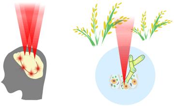
This is an illustration of optical stimulation of cell activity in human brain and plant cells by holographic optogenetics. Courtesy of Kobe University.
Professor Osamu Matoba said, “We have a research grant from JST CREST Grant Number JPMJCR1755, Japan, to fabricate a SIFOM and then apply it to further development of neuroscience. We will collaborate with companies to introduce the new optical microscope into the commercial market.”
The research was published in
Optics Letters, a publication of OSA, The Optical Society (
https://doi.org/10.1364/OL.43.005447).