Physicists at Bielefeld and Frankfurt Universities are studying blood cells, algae and bacteria by trapping these biological cells with a laser beam. Using this procedure they have obtained superresolution images of the DNA in single bacteria.
Professor Thomas Huser, head of the Biomolecular Photonics Research Group in the Faculty of Physics, said their technique offers a different way to examine cells and does not change the cells in the preparatory stage.
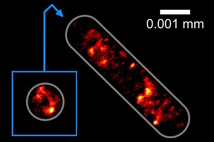
Picture of the
distribution of the genetic information in an Escherichia coli bacterial cell:
Physicists at Bielefeld University are the first to photograph this
distribution at the highest optical resolution without anchoring the cells on a
glass substrate. Courtesy of Bielefeld University.
“Our new method enables us to take cells that cannot be anchored on surfaces and then use an optical trap to study them at a very high resolution,” said Huser. “The cells are held in place by a kind of optical tractor beam. The principle underlying this laser beam is similar to the concept to be found in the television series Star Trek.”
The futuristic technique also allows the specimens to be turned and rotated, and the laser beam functions as an extended hand for making small adjustments microscopically.
The Bielefeld and Frankfurt team has further developed the procedure for use in superresolution fluorescence microscopy, a process that was once only possible with electron microscopy, where a laser beam is directed toward the cells to cause them to light up.
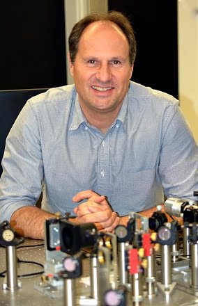
Prof. Dr.
Thomas Huser and his team have further developed a procedure for the
superresolution microscopy of cells. Courtesy of Bielefeld University.
Robin Diekmann, a member of the Biomolecular Photonics Research Group, said in their new method a second laser beam is used as an optical trap causing the cells to float under the microscope.
“The laser beam is very intensive but invisible to the naked eye because it uses infrared light,” said Diekmann. “When this laser beam is directed towards a cell, forces develop within the cell that hold it within the focus of the beam.”
Using their new method, the Bielefeld physicists have succeeded in holding and rotating bacterial cells in such a way that they can obtain images of the cells from several sides. Thanks to the rotation, the researchers can study the three-dimensional structure of the DNA at a resolution of circa 0.0001 millimeters.
Professor Huser and his team plan to further modify their method to enable the observations of interplay between living cells.
The study was published in the journal Nature Communications (doi:10.1038/ncomms13711).