
Optics Peers Through the Haze of Dementia
Imaging of the eyes is providing a window into the brain deterioration of those with Alzheimer’s disease, promising early diagnosis and potentially benefiting both patients and researchers.
As the number of people suffering from Alzheimer’s disease (AD) and related dementias grows, so too does the pressure on the medical community to identify dementias as early as possible and potentially treat them. Researchers on both sides of the Atlantic are using optical technologies to capture the manifestations of dementia in the brain before outward symptoms of the condition occur.

Optics may hold the key to unlocking the mysteries of memory loss and AD.
And as it turns out, those with an eye toward early AD diagnosis may just have found the answer in the eyes of people experiencing its beginning stages. At Duke University, researchers are using multimodal retinal imaging — including OCT, OCT angiography, ultrawide-field color photos, and ultrawide-field autofluorescence images, among other methods — to examine levels of degeneration in the retinas of people with AD (Figure 1). Ophthalmologists have known for many years the connection between AD and the thinning of retinal tissue and the reduction in vein density1.

Figure 1. Duke University researchers look into the eyes of their subjects for warning signs of AD. Courtesy of Duke Eye Center.
Duke researchers Dilraj S. Grewal, M.D., Bryce W. Polascik, Sharon Fekrat, M.D., and others, began an inquiry with a case study of 96-year-old female identical twins, one with AD and one without, through the use of OCT angiography (OCTA) imaging. OCTA is a noninvasive approach that can visualize blood vessels with diameters as small as 5 µm.
According to researchers, both twins had received cataract surgery treatement at the time of the study. The cognitively normal twin had diagnoses of Fuchs’ dystrophy (which leads to swelling of the cornea) and geographic atrophy caused by nonneovascular age-related macular degeneration (AMD) in both eyes. The twin with AD also had Fuchs’ dystrophy, geographic atrophy caused by nonneovascular AMD in both eyes, as well as a history of a retinal detachment in the right eye, repaired by vitrectomy and scleral buckling surgery2.
With OCTA (6- × 6-mm scan), the team observed a notable difference in the twins’ superficial vascular plexus (SVP). The SVP is supplied by the central retinal artery and composed of larger arteries, arterioles, capillaries, venules, and veins. The twin with AD showed a loss of vessel density in the macula, along with a decreased subfoveal choroidal thickness (75 µm vs. 114 µm in the twin without AD). The choroid is a vascular tissue deep in the retina that provides oxygen and nourishment to the outer portion of the retina and its photoreceptors. The fovea centralis is a small, central pit composed of closely packed cones in the eye. Figure 2, though not drawn from the example of the twins, illustrates this phenomenon of deterioration.
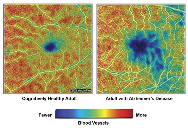
Figure 2. Images from a Duke University study show the loss of blood vessels in the retina of an adult with AD (right) versus an adult with a healthy brain. Courtesy of Duke Eye Center.
A continental vision for change
In Europe, the MOON (Multimodal Optical diagnosis of Ocular and Neurodegenerative disease) project — funded by the EU and involving collaborations with the Medical University of Vienna (Med-Uni Vienna), the Netherlands Organisation for Applied Scientific Research, and the Leibniz Institute of Photonic Technology — is proceeding along a similar path toward detection of AD in the eyes. Rainer Leitgeb, an associate professor at MedUni Vienna and the coordinator of the project, said the focus — literally and figuratively — has been on the recognition of molecular biomarkers, or measurable substances that indicate the presence of an illness or condition, through the use of Raman spectroscopy. Leitgeb said the MOON consortium believes that Raman spectroscopy, which measures vibrational modes of molecules, provides the greatest specificity at the molecular level.
“Our study aims at a noninvasive diagnosis of diseases, including … Alzheimer’s. We left out, therefore, invasive blood tests, or tests of the cerebrospinal fluid,” Leitgeb said. “We also concentrated on optical technologies, as they provide high resolution, combined with high molecular specificity, and can be easily and noninvasively applied to the human eye. The key assumption is that the eye serves as a window to the brain, and, as such, to pathologic changes due to neurodegenerative diseases” (Figure 3).
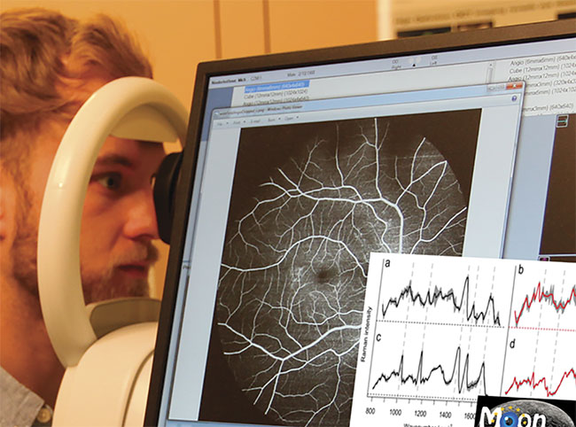
Figure 3. The use of various technologies to identify neurodegeneration through examination of the
eyes is the charge of the MOON project in Europe. Courtesy of the MOON consortium.
Agencies follow research closely
Heather Snyder, vice president of medical science relations at the Alzheimer’s Association, believes these discussions are timely, given the growing epidemic. The nonprofit organization reports that as of this year, 5.8 million Americans live with AD, which is characterized by a progressive loss of cognitive functioning, the shrinking and destruction of brain cells, and, ultimately, death. This number is expected to rise to 14 million by 2050. AD is the sixth leading cause of death in the U.S. and is expected to cost the nation some $290 billion in health or other services this year (Figure 4).
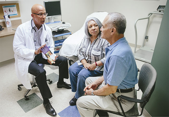
Figure 4. A couple consults with a doctor about living with symptoms of AD. Courtesy of the
Alzheimer’s Association.
The Alzheimer’s Association shows no preference for one form of technology over another. Snyder said she was aware, however, that several years ago, a number of studies presented at the Alzheimer’s Association International Conference confirmed that changes in the eye were congruent with changes in the brain itself. “We are looking for as much research as we can get through technologies like OCT and OCTA and whatever other imaging will give us a picture of changes of blood flow or other warning signs of what is occurring,” she said. “Independent research groups will go forward, and when the science tells us what is needed, we will support it.”
The Alzheimer’s Association isn’t the only agency with an eye toward development of research. Much of the work in the academic realm is funded nationally through grants from the National Institute of Biomedical Imaging and Bioengineering (NIBIB) of the National Institutes of Health (NIH). The stated mission of NIBIB is to fund research for the development of new biomedical imaging and bioengineering techniques and devices to fundamentally improve the detection, treatment, and prevention of disease.
“We aim to fund imaging and interventions for any stage of the disease, as well as early-stage detection methods,” said Randy King, NIBIB program director for ultrasound, optical imaging, and spectroscopy in the Division of Applied Science and Technology. He explained that first a proposal is considered by a scientific review group composed of nonfederally supported scientists in a particular field, who judge the likely impact of the research on the field in question. Then a potential project moves on for consideration by advisory councils and/or review boards, which determine the technical merit of the priorities in a study.
King said one NIH initiative related
to AD that the NIBIB funded and participated in was a collaboration with the National Institute on Aging. This project directed “investigators to request administrative supplements to develop new tools, technologies, and approaches for the prevention, diagnosis, treatment,
and understanding of Alzheimer’s disease and Alzheimer’s disease-related dementias.” Technologies being applied
or considered include the use of near-infrared light to sense intracranial pressure (Carnegie Mellon University) and acousto-optic modulated interferometric diffuse correlation spectroscopy (iDCS), which measures cerebral blood flow, at 1064 nm (Massachusetts General Hospital).
A focus on those at risk
The NIH and NIBIB have also funded the development of a high-speed optical imaging system to be used in the Alzheimer’s Disease Research Center (ADRC) at the University of California, Irvine (UCI). According to Yama Akbari, M.D., an assistant professor in the departments of neurology and neurological surgery at UCI, the system uses laser speckle imaging in combination with spatial frequency domain imaging (SFDI) to monitor the cerebral blood flow and brain metabolism in live animals (Figure 5). Akbari is working with an interdisciplinary team that includes professor Bernard Choi, postdoctoral fellow Christian Crouzet, and project scientist Robert Wilson.
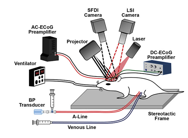
Figure 5. A high-speed optical imaging system in use at the University of California, Irvine to
image cognitive activity and decline in live mice. LSI: laser speckle imaging; BP: blood pressure;
AC-ECoG: alternating current electrocorticography; DC-ECoG: direct current electrocorticography.
Courtesy of Christian Crouzet.
“Using the optical imaging system, we are determining the impact of aging and clinical events, such as hypoxia and sleep apnea, on brain hemodynamic and functional changes in a sporadic AD mouse model,” Akbari said. “More specifically, we are using our multimodal optical imaging system to determine how these perturbations affect CBF [cerebral blood flow], brain metabolism, neurovascular coupling, and pulsatile factors in AD and control mice at various ages.”
Akbari credited professor Bruce Tromberg, director of the NIBIB, with co-inventing and pioneering the SFDI system at the UCI Beckman Laser Institute. The Clarifi Imaging System, which received FDA approval last year, uses SFDI, which is based on structured light, to help clinicians and researchers identify tissue function and compromised circulation. This can aid in the early diagnosis of certain conditions.
Akbari said that with this technology, researchers can get fast measurements of the cerebral metabolic rate of oxygen consumption, which he described as “one of the holy grails of brain metabolism/function and often not easy to measure.” He then went on to explain that the NIH funding enables the investigation of cerebral blood flow and brain metabolism with resolution on the order of milliseconds.
“Currently, our work is mostly investigative, to see how AD mice have altered CBF and brain metabolism, using our fast optical imaging system. And our preliminary data does point towards changes in CBF and brain metabolism,” Akbari said. “The next logical step for us is to do more thorough studies and then assess maneuvers to counteract any pathological changes during treatment paradigms. There are indeed ways that we can alter CBF and brain metabolism, and we hope to test these types of treatments in the future.”
References
1. F. Berisha et al. (May 2007). Retinal abnormalities in early Alzheimer’s disease. IOVS, Vol. 48, Issue 5, pp. 2285-2289.
2. D.S. Grewalet al. (June 2018). Assessment of differences in retinal microvasculature using OCT angiography in Alzheimer’s disease: a twin discordance report. OSLI Retina, Vol. 49, Issue 6, pp. 440-444.
A Treatment of Sight and Sound
Researchers at MIT are gauging whether optics could be used to
not only detect Alzheimer’s disease but to treat it as well.
In recently published research in Neuron, authors from MIT and the RIKEN Center for Brain Science noted, “Human AD patients show reduced power of oscillations in the gamma frequency band (30-120 Hz)1.” The researchers explained that, based on their findings with studying mouse models of AD, flickering light at 40 Hz (using a custom LED device) over a period of days significantly reduced the telltale amyloid plaque levels and neuroinflammation that interfere with neural synapses and circuit function. They saw evidence, in comparison to a control group, that after the light exposure, the activity and gene expression signatures inherent in some brain cells — whether individually or collectively — is restored for an extended period of time, and learning and memory were improved.
“We are extending our approach to other disease indications,” said Li-Huei Tsai, director of the Picower Institute for Learning and Memory at MIT, and a leader of this research. “We are also exploring the mechanisms by which our approach provides neuroprotection.”
Tsai co-founded Cognito Therapeutics Inc. to commercialize the therapies that were in use in the university setting. Zach Malchano, president of the company, said that, while he couldn’t reveal all aspects of the technology in use, developers envisioned a wearable device people could use on a schedule in their own homes.
“What we are moving toward [are] noninvasive means that use sensory pathways, both visual and auditory,” he said. “The blood/brain barrier is always a difficult challenge for treatment, and effects can be off target. So we have focused on sensory stimulation. We see our approach as long-term therapy that is accessible and fits in with patients’ lives.”
Malchano said three clinical trials of the technology are underway, and recruitment of volunteers has begun.
Some of the research team’s findings will be presented in a special lecture by Tsai at Neuroscience 2019, to be held by the Society for Neuroscience in Chicago this month.
Reference
1. C. Adaikkan et al. (June 2019). Gamma entrainment binds higher-order brain regions and offers neuroprotection. Neuron, Vol. 102, Issue 5, pp. 929-943.
Published: September 2019