
Optofluidic Microscope Invented
PASADENA, Calif., Sept. 11, 2006 -- The old optical microscopes that everyone used in high school biology class may be a step closer to the glass heap. Researchers at the California Institute of Technology have announced their invention of an optofluidic microscope that uses no lens elements and could revolutionize the diagnosis of certain diseases such as malaria.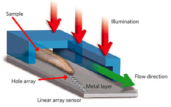
The optofluidic microscope, and the implementation scheme for an on-chip optofluidic microscopy. The device is uniformly illuminated from the top. The target sample flows through the channel, and the transmission through each hole is acquired and recorded. The composition of the transmission traces creates a transmission image of the target sample. (Images: Nature)
Reporting in the journal Lab on a Chip, Caltech assistant professor of electrical engineering professor Changhuei Yang and his coauthors describe the novel device that combines chip technology with microfluidics. Although similar in resolution and magnifying power to a conventional top-quality optical microscope, the optofluidic microscope chip is only the size of a quarter, and the entire device -- imaging screen and all -- will be about the size of an iPod.
"This is a new way of doing microscopy," said Yang, who also has a dual appointment in bioengineering at Caltech. "Its imaging principle is similar to the way we see floaters in our eyes. If you can see it in a conventional microscope and it can flow in a microfluidic channel, we can image it with this tiny chip."
That list of target objects includes many pathogens that are most dangerous to human life and health, including the organism that causes malaria. The typical method of diagnosing malaria is to draw a blood sample and send it to a lab where the sample can be inspected for malaria parasites. A high-powered optical microscope with lens elements is far too big and cumbersome for inspection of samples in the field.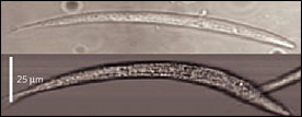
A conventional microscope image of Caenorhabditis elegans (top) and an optofluidic microscope (500-nm resolution limit) image, also of C. elegans.
With a palm-sized optofluidic microscope, however, a doctor would be able to draw a drop of blood from the patient and analyze it immediately. This process would be much simpler and faster than the current method, and the equipment would be far cheaper and more readily available to physicians in third-world countries.
The device works by literally flowing a target sample across a tiny fluid pathway. Normally, the image would be low in resolution because the target would interrupt the light on a single pixel, thus limiting the resolution to pixel size.
However, the researchers have avoided this limitation by attaching an opaque metal film to a microfluidic chip. The film contains an etched array of submicron apertures that are spaced in such a way that adjacent line scans overlap and all parts of the target are imaged.
The new optofluidic microscope is one of the first major accomplishments to come out of Caltech's Center for Optofluidic Integration, which was begun in 2004 with funding from DARPA for development of a new generation of small-scale, highly adaptable and innovative optical devices.
"The basic idea of the center is to build optical devices for imaging, fiber optics, communications, and other applications, and to transcend some of the limitations of optical devices made out of traditional materials like glass," said Demetri Psaltis, who is the Myers Professor of Electrical Engineering at Caltech and a coauthor of the paper. "This is probably the most important result so far showing how we can build very unique devices that can have a broad impact."
Xin Heng, a graduate student in electrical engineering at Caltech, performed most of the experiments reported in the paper. The other Caltech authors are David Erickson, a former postdoctoral scholar who is now a mechanical-engineering professor at Cornell University; L. Ryan Baugh, a postdoctoral scholar in biology; Zahid Yaqoob, a postdoctoral scholar in electrical engineering; and Paul W. Sternberg, the Morgan Professor of Biology. The work was also reported in the journal Nature in July.
For more information, visit: www.caltech.edu
Published: September 2006