A novel optogenetics platform, dubbed optoDroplet, uses light to activate phase transitions inside living cells. The optoDroplet system is being used to study the condensed phases driven by intrinsically disordered protein regions (IDRs). Its use could lead to a better understanding of cellular processes, including how proteins assemble and the potential link between protein aggregates and disease.
Researchers at Princeton University used the optoDroplet system to induce phase transitions in cells driven by the IDRs of various ribonucleoproteins (RNPs), including FUS, DDX4 and HNRNPA1. Phase transitions in cells were induced and controlled by increasing or decreasing the intensity of light, and level of light intensity was recorded.
Protein droplets appear as white dots as a cell is exposed to light from a laser. Courtesy of Princeton University.
Using mouse and human cells, the team spliced in a gene for a light-sensitive protein from a mouse-ear cress plant (Arabidopsis thaliana). The light-sensitive tag was fused to protein components believed to drive phase transitions in living cells. When exposed to blue light, the proteins mimicked the condensation process that naturally occurs in cells.
"To use the analogy of water vapor, you can think of what we did as using a laser to locally change the temperature of some area of the air so that water droplets would condense out of it," said professor Clifford Brangwynne, who was the senior author of the paper on the research.
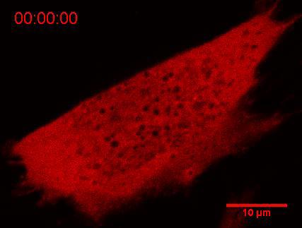
Protein droplets appear as white dots as a cell is exposed to light from a laser. Courtesy of Princeton University.
The team repeatedly prompted the proteins to condense and then dissolve by turning the light on and off. The process proved fully reversible, even after many activation cycles. However, cells exposed to high-intensity light or high concentrations of proteins formed semi-solid gels that, while initially reversible, over time solidified into aggregates similar to those associated with some diseases.
The researchers were able to determine when condensed liquid protein droplets formed, as well as when solid-like protein aggregates formed. The optoDroplet system thus elucidated not only physiological phase transitions but also their link to potentially pathological aggregates.
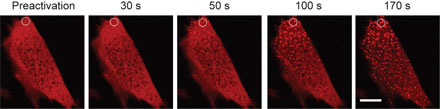
Protein droplets, appearing as white dots, increase in number as intensity of a laser shining on the cell increases. Courtesy of Princeton University.
"OptoDroplet provides us a level of control we can use to precisely map what we call the phase diagram in living cells," said Brangwynne. "With that, we're beginning to understand how cells use their natural machinery to move through this intracellular phase diagram to assemble different types of organelles.
"We've shown with optoDroplet that we can readily assemble and disassemble phase-separated liquids, and they do not appear to cause any problem for the cell," Brangwynne added. "But the gel-like assemblies appear to be more problematic, since over many cycles, they develop into persistent aggregates that the cell can no longer deal with and that can start to gum up healthy biological processes."
To date, most studies have used purified proteins studied in test tubes; and researchers have had few methods to study phase transitions in living cells. The optoDroplet system could help advance scientists’ understanding of when phase transitions go awry.
"This optoDroplet tool is starting to allow us to dissect the rules of physics and chemistry that govern the self-assembly of membraneless organelles," said Brangwynne. "The basic mechanisms underlying this process are very poorly understood, and if we get a handle on it, there might be a hope for developing interventions and treatments for devastating diseases connected with protein aggregation, such as ALS."
Edward Lemke, a researcher at the European Molecular Biology Laboratory in Heidelberg, who was not involved in the study, said, "The proteins targeted by optoDroplet are an important constituent of phase-separating proteins, many of which are also associated with infamous diseases. The optoDroplet system gives access to modulating the state of these proteins inside the cell in a minimally invasive and highly controlled fashion, so it can provide new insights on how they carry out their function."
The research team will continue to experiment with optoDroplet to better understand the behaviors of living cells.
"This is fundamental science we're doing, answering basic questions about phase transitions in cells," Brangwynne said. "But we're hoping these insights will reveal not only how healthy cells work, but also how they can become diseased, and maybe eventually cured."
The research was published in Cell (doi: 10.1016/j.cell.2016.11.054).