Photodynamic therapy could be the future of cancer treatment – that is, once the photoactive chemicals behind it start doing their jobs better.
Photodynamic therapy (PDT) is proving to be a more than viable option for cancer treatment. Compared with other treatments, such as chemotherapy and radiation therapy, PDT is more selective, causing far less damage to healthy cells near cancerous targets due to the precise way in which photosensitizers can locate and infiltrate tumor cells.
The task remains to find the best combinations of photosensitizers and conjugates to propel the technique past chemotherapy and other traditional methods.
Basically, a photoreactive chemical compound called a photosensitizer is introduced into a patient, where it aggregates near an active tumor site. A clinician then shines light from a diode laser or LED source onto the tumor region. The light, which has a specific wavelength, activates the photosensitizer without affecting surrounding healthy tissue. Once activated, the photosensitizer transfers some of its energy to nearby ground-state molecular oxygen, producing excited singlet oxygen. The result is oxidation of tumor cells in the site, destroying the cancer while harming as few of the adjacent healthy cells as possible.
Bringing light to the site is an ongoing issue. Early studies of PDT focused on skin cancers, such as various types of melanoma, because it was easier to shine near-infrared wavelengths a couple of millimeters through the skin’s surface to the tumor site. This drove design of the first photosensitizers to favor compounds that would preferentially react to light in that range. More recently, advances in endoscopic light-delivery systems have made deeper tumors easier to reach and have broadened the range of potential wavelengths and matching photosensitizers.
Different compounds are now used as photosensitizers, including phthalocyanine, chlorine, bacteriochlorin and porphyrin. None, however, is a perfect candidate.
Getting a photosensitizer to find and attach itself to a tumor cell is a major battle. The body’s immune system, for example, seeks out and annihilates some forms of photosensitizers, reducing the effectiveness of the overall treatment. Adding antibodies to photosensitizers can help their affinity for cancer cells, but some researchers feel that protecting them with shells composed of lipoproteins is a better way to go. Lipoproteins not only help their cargo locate and infiltrate tumors, but also help protect them from enzymes and macrophages that might alter or destroy them before they even arrive at the treatment site.
Gold nanoparticles and liposomes also have been considered as adjuncts that could help photosensitizers directly enter cancer cells.
Gold rush
Researchers at Rhodes University in Grahamstown and in the biophotonics department of the National Laser Center in Pretoria, both in South Africa, are among the groups looking at the possible improvements to photosensitizer action provided by gold nanoparticles.
Tebello Nyokong of Rhodes University and her colleagues had gold nanoparticles in mind during efficiency tests of a particular photosensitizer, as they reported in the Feb. 6 issue of the Journal of Photochemistry and Photobiology B: Biology.
Phthalocyanine compounds strongly absorb in the 600- to 800-nm range, yet tissues are transparent to a useful degree to these wavelengths. The result is an ability to reach deeply into tissue and provide sufficient energy to the photosensitizer to activate it.
Several attributes must be considered when designing a photosensitizer, said Nyokong, director of the Nanotechnology Innovation Center at Rhodes. One is that it should have a high specificity for cancer, which is achieved through inclusion and coordination of molecules such as folic acid and vitamin B12. Another highly valued attribute is good absorption in the red wavelengths, which is aided by sulfur linkages in the photosensitizer compound. The final product also should be water-soluble and initiate large production of singlet oxygen, which drives tumor cell death.
The group’s candidate was [2,9,17,23-tetrakis-(1,6-hexanedithiol)phthalocyaninato]zinc(II), a second-generation phthalocyanine-based compound. Its target: human malignant breast cancer cells (MCF-7).
The researchers chose zinc over more typical sulfur in their compound because it enhances the production of singlet oxygen while being somewhat easier to assemble. After forming the phthalocyanine complexes, they introduced some to gold nanoparticles, which self-assembled with the compound. Others were bound to liposomes as a delivery vehicle.
Using a Shimadzu spectrophotometer, a Varian Inc. spectrofluorimeter, and a single-photon counter and diode laser made by PicoQuant GmbH, the investigators measured the absorption spectra, fluorescence excitation and fluorescence lifetimes of their photosensitizer in action against MCF-7.
After determining that a light dose of 4.5 J/cm2 provided adequate intensity without harming nearby healthy cells, the researchers compared how well the gold nanoparticles and the liposomes aided the overall phototoxic effect.
They found that, after photoactivation of the two complexes, 60.1 percent of the tumor cells treated with nanoparticle-enhanced phthalocyanine remained viable, whereas the liposome-enhanced complexes fared much better with 51.9 percent cell viability.
Nyokong’s work with PDT is focused on synthesizing bifunctional agents – compounds that serve two functions, generally enhancing location and attachment to tumor cells. In her lab, the desired result is agents that combine the action of PDT and other treatments, such as hyperthermia (destroying tumors with applied heat, which increases the uptake of oxygen, thus accelerating cell destruction).
Nyokong’s lab also is looking at combinations of chemotherapy and PDT via introduction of platinum to common photosensitizers. Next up for her group is the ongoing search for water-soluble phthalocyanine compounds that include liposomes. Better water solubility, the researchers say, should improve the ability of phthalocyanine to generate singlet oxygen inside cells.
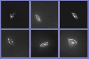
Before and after images show the effect of a porphyrin-based photosensitizer that was carried into HeLa cells by a ruthenium-based, cube-shaped cage. Courtesy of the Journal of the American Chemical Society.
Cage death matches
Phthalocyanine- and porphyrin-based photosensitizers struggle to reach the tumor site because they are fairly poorly water soluble. Placing either type of complex inside the hydrophobic cavity of an otherwise water-soluble vessel designed to wend its way breezily toward target cancer cells is thought by several research groups potentially to improve the situation.
The tricky part is getting the vessel to unload its cargo upon arrival.
Another way to bring the photosensitizer to the cell is to wrap it inside an organometallic cage. This helps address the water-solubility issue while offering control of photosensitizer release, according to Bruno Therrien, an associate professor at the University of Neuchâtel in Switzerland.
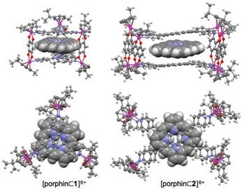
Side and top views show 3-D models of prism-shaped (left) and cubic (right) “metalla-cages” designed to transport photosensitizers to tumor cells. Courtesy of the Journal of the American Chemical Society.
Therrien and his colleagues at the university and at Centre Hospitalier Vaudois in Lausanne, Switzerland, devised and tested two types of carrier vessels to ferry porphyrin to its target. One, in the form of a prism, locks the porphyrin tightly; the other, a cubelike structure, is a more flexible jacket. Both vessels are made with ruthenium-based compounds.
“Within the ‘metalla-prism,’ it’s a ship-in-the-bottle system – only breakage of the cage can release the guest,” Therrien said. “However, from the ‘metalla-cube,’ the porphyrin is free to go through one of the apertures without [breaking] the cage.”
With either vessel, the porphyrin remains unreactive to light and only becomes photosensitive once released.
The group studied the uptake of both vessel types and their cargo into HeLa cells, and then used a 488-nm laser made by Spectra-Physics at various doses to release, then activate, the porphyrin once the metalla-cages were inside the cell membranes.
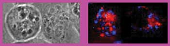
Fluorescence micrographs of HeLa cells show how the photosensitizer chlorin e6 (top row) and a complex of chlorin e6 and poly-L-lysine (bottom row) accumulate inside HeLa cells after 10 min (left), 1 h (center) and 2 h (right). Note how the photosensitizer alone remains in the cytoplasm near the cell membrane, while the conjugated pair works its way from the inner wall to the cell nucleus. Courtesy of Current Topics in Medicinal Chemistry.
Once released, porphyrin discharged from either cage performed well at generating singlet oxygen and thus destroying the HeLa cells. Interestingly, the porphyrin delivered via the cubelike metalla-cages packed more punch, requiring one-tenth the energy (0.2 J/cm2) to reach the threshold where half the cells are killed compared with the metalla-prism combo (2.1 J/cm2).
Both controlled release of the photosensitizer and its ultimate phototoxicity are important, according to Therrien. “Controlled release can eliminate side effects, such as skin photosensitivity after and during treatment, but the active treatment is phototoxicity, so you still need an efficient photosensitizer.”
The ultimate goal of Therrien and his colleagues is to be able to irradiate at a specific wavelength to break up the cage where and when it is desired and, after release, apply a second dose of light to activate the photosensitizer.
Location, location, location
One of the most troubling disadvantages of first- and second-generation photosensitizers, according to researchers at the Tokyo Institute of Technology, is that they do not locate cancer cells as well as they might. The more specifically diseased cells are targeted, the more healthier viable cells can remain.
“(A) photosensitizer which shows high tumor localization shows low phototoxicity for normal tissue,” said Shun-Ichiro Ogura of the institute’s department of biomolecular engineering. “It is quite important for tumor therapy.”
But as importantly, the short lifetime of singlet oxygen (measured in no more than microseconds) means that the closer they are to the right target, the more damage they can do. Therefore, improving localization can improve PDT efficacy.
Some photosensitizers, such as porphyrin-based constructions, accumulate in a target cell’s plasma membrane. However, the nucleus is the place to be if you want maximum destructive impact. Ogura and his colleagues found that one possible way to get to the cell nucleus effectively is to combine the popular photosensitizer chlorin e6 with poly-L-lysine. By itself, chlorin e6 stays in the cytoplasm of the cell, but the conjugated pair ultimately travels to the nucleus. After subsequent light exposure, the complex offered high phototoxicity.
The group presents its findings on the localization capabilities of several photosensitizer types in the February issue of Current Topics in Medicinal Chemistry.