Hybrid polymer solar cells are not as common as silicon cells, nor are the efficiencies as high – currently only about 2 percent. But hybrid polymer cells, which are both organic and inorganic, offer two major advantages that keep researchers seeking ways to boost their efficiency. Most notably, they can be printed in a roll-to-roll process, reducing the cost of their manufacture, and they’re flexible and can be applied to a range of objects such as clothes, umbrellas, laptop carrying cases and more.
Hybrid cells are a mixture of a polymer and a metal oxide, a combination that creates charges when the mixture soaks up the sun. Critical to the cells’ efficiency is the degree of mixing, which can enhance the area of the interface where the charges are formed. Researchers at the Eindhoven University of Technology in the Netherlands and at the University of Ulm in Germany used 3-D transmission electron microtomography to peer inside these solar cells. They are interested in their structure because it’s difficult to control, as a result of the differences between the chemical nature of polymers and metal oxides. The researchers used a precursor compound that mixes with the polymer and then converts into the metal oxide only after is incorporated in the photoactive layer.
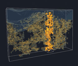
This 3-D electron tomography image of the inside of a polymer-metal oxide solar cell shows the metal oxide network (yellow) in the polymer phase (transparent) below an aluminum top electrode (gray). Most zinc oxide is interconnected and makes contact with the aluminum electrode.
A commercial 3-D transmission electron microscope (TEM) from FEI Co., a Tecnai G2 20, was used to visualize the modules. Lead researcher René Janssen, a professor of molecular materials and nanosystems, noted that this is the first time that 3-D tomography has been applied to hybrid solar cells. He said its application is similar to that of a medical CT scanner in that researchers look at the transmission of an electron beam through the sample and take images while rotating it in 1° steps. A series of 2-D TEM projections is then used as input to come up with a 3-D reconstruction, which gives insight into the morphology.
Measuring percolation
“There are quite a few of these bulk heterojunction solar cells, but so far we have very little quantitative information on the morphology. By using 3-D tomography, we have a way to understand how these devices work,” he explained.
In general, the challenge is to be careful that the electron beam doesn’t influence the sample or damage the very thin film, according to Janssen. However, because of the large difference in density between the polymer and the zinc oxide, the resulting images showed good contrast and demonstrated in three dimensions the importance of mixing.
Researchers from the Institute for Stochastics at the University of Ulm extracted the typical distances between the components that relate to the efficiency of charge formation. Then they analyzed how much of each component is connected to the electrode for charge transport. Through this research, they were able to analyze and quantify the percolation pathways. The next step will be to use this data to develop a detailed operational model of these cells, which they hope will help solar researchers better understand the way organic and inorganic solar cells work.