A research team led by Lihong Wang of the California Institute of Technology has developed a two-dimensional multifocal optical resolution photoacoustic microscopy (MFOR-PAM) system. The team used a 2D microlens array for optical excitation, and an acoustic ergodic relay to simultaneously detect the photoacoustic (PA) responses to the multifocal optical illuminations with a single element ultrasonic transducer.
The system, referred to as multifocal optical-resolution photoacoustic microscopy through an ergodic relay (MFOR-PAMER), shortened the scanning time by at least 400× compared to conventional OR-PAM systems at the same imaging resolution, while maintaining a simple and economic setup.
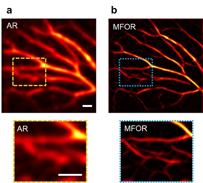
(a) AR-PAMER image. (b) MFOR-PAMER image. AR, acoustic resolution; MFOR, multifocal optical resolution; All scale bars, 1 mm. Courtesy of Caltech.
The novel MFOR-PAM system is centered around a key enabling element known as the acoustic ergodic relay. For photoacoustic imaging an ergodic relay — such as a light-transparent prism — can be used as an encoder to transform PA signals from different input positions into unique temporal signals. By recording the system impulse response of each input position in advance, the PA signals from the entire field of view can be detected in parallel upon a single laser shot. Then, the encoded PA signals can be decoded mathematically to reconstruct a 2D-projection image of the object.
The system uses a microlens array to focus a wide-field laser beam into multiple optical focal points. Unlike a conventional focusing lens that needs to scan a single optical point across the entire field of view, the microlens array can reduce the time required to form an image by scanning multiple spots at once.
According to the researchers, the system has potential for numerous biomedical applications, such as UV illumination for high-speed, label-free histological study of biological tissues. The design can reduce imaging time from several hours to less than a minute, making its application more practical.
The research was published in Light: Science & Applications (www.doi.org/10.1038/s41377-020-00372-x).