While traditional optical coherence tomography produces motion artifacts due to insufficient imaging speed, photonic integrated circuit-based systems could enable high-speed imaging by scanning multiple points on the eye at once.
By Aaron Adkins, Senyue Hao, and Chao Zhou
In the U.S., eye diseases and vision problems are widespread, with diseases such as age-related macular degeneration (AMD), glaucoma, and diabetic retinopathy affecting millions of people. The NIH estimates that by 2030, 5 million Americans will have low vision, and 2.2 million will be blind, and the total economic burden for vision-related issues has been estimated to be $139 billion1. Advanced diagnostic methods aimed at early and effective identification of such diseases, including some of the most recent iterations of optical coherence tomography (OCT), hold great potential to allow for early treatment and to prevent permanent vision issues.

A cross-sectional view of the eye obtained via OCT. Courtesy of iStock.com/yogenyogeny.
OCT is one of the most powerful diagnostic tools used by modern ophthalmologists, providing high-resolution 3D images of the interior structure of the retina and anterior segment of the eye (opening image). OCT traces its origins to a seminal 1991 paper from a group at MIT led by professor James G. Fujimoto2. Shortly after its introduction, OCT was rapidly adopted and commercialized for ophthalmic imaging, with the first OCT instruments becoming available in 1996.
Since its inception, OCT has undergone technological advancements in instrumentation and performance and has solidified itself as a standard for eye disease diagnosis. As a testament to the enormous impact of OCT, pioneers of OCT technology — David Huang, Eric Swanson, and James G. Fujimoto — were awarded the Lasker-DeBakey Clinical Medical Research Award and the National Medal of Technology and Innovation in 2023 for their invention of OCT and their contributions to ophthalmology3.
OCT principles and challenges
OCT is fundamentally based on the principle of low-coherence interferometry. Light from a wide-bandwidth laser source is split into two paths, with one path reflecting off a reference mirror, and the other path being focused onto the imaging sample — for example, the retina. The imaging beam passes through the transparent vitreous fluid and penetrates the retinal tissue, scattering off multiple retinal layers with varying intensities. This backscattered light is then recombined with the reflected light from the reference mirror, forming an interference pattern from which a depth profile of the sample can be computed. This depth profile is called an A-scan. By laterally scanning this beam across the sample, depth profiles can be obtained across a 2D area, forming a 3D image.
Since its inception, OCT has undergone technological advancements in instrumentation and performance and has solidified itself as a standard for eye disease diagnosis.
While OCT has been widely adopted in clinical ophthalmology, it still suffers from a few limitations that hinder its full potential as a diagnostic tool. Modern OCT devices can perform a full scan of the retina in <10 s, but this is still too slow to eliminate motion artifacts caused by involuntary eye movements during data acquisition. Ideally, an OCT scan could be performed in <1 s, eliminating artifacts introduced by microsaccades, involuntary movements of the eye occurring 1 to 2×/s. Current solutions to this issue include active motion correction and image registration with fundus photography, but the most straightforward solution would be to increase the imaging speed. However, simply boosting the speed leads to decreased signal-to-noise ratio, as the imaging beam spends less time on each sample point, limiting the amount of light that can be collected back from the sample.
Space-division multiplexing OCT
An elegant approach to effectively increase OCT speed is to parallelize the imaging across multiple beams, each scanning a separate physical location on the sample. This method, called space-division multiplexing OCT (SDM-OCT), was pioneered by the authors’ group at Lehigh University and Washington University in St. Louis4. The architecture of an SDM-OCT system consists of a standard swept-source OCT system, with the so-called “sample arm,” which focuses on layers of the eye, optically split into multiple parallel channels. These channels each scan a small section of the total region of interest on the sample. The beams are recombined into the system, with optical delay introduced to prevent overlap of the signals. A single photodetector then acquires the separate images from all parallel beams.
The individual signals are then stitched together into a full image of the sample. In this manner, the imaging speed is directly boosted by a factor equal to the number of parallel beams. Imaging speed in OCT is quantified as A-scans per second, with current commercial systems typically operating at around 50,000 to 100,000 A-scans/s. A proof-of-principle four-channel SDM-OCT was previously constructed with off-the-shelf fiber optic components and was able to image the retina at an effective speed of 800,000 A-scans/s (Figure 1)5.

Figure 1. A space-division multiplexing OCT (SDM-OCT) retinal imaging of a healthy volunteer (a). An en face projection of the retinal pigment epithelium (RPE) layer (b), with the yellow line indicating the location of the vertical cross section (c) and the green line showing the location of the horizontal cross section (d) (CHR: choroid; EZ: ellipsoid zone; ILM: internal limiting membrane; IPL: inner plexiform layer; ONH: optic nerve head; OPL: outer plexiform layer). Retinal thickness map (e). A rendering of the 3D OCT data set of the entire retina acquired in 1 s (f). Scale bars = 1 mm. Adapted with permission from Reference 5.
Despite the dramatic performance improvement of SDM-OCT in this proof of concept, the construction of a purely fiber-based system for clinical use is complex, requiring precise splitting and splicing of fibers that necessitates a high degree of manual labor. The difficulty is compounded with higher numbers of parallel channels, limiting scalability. Based on direct experience constructing the four-channel fiber-based system, the authors found that these challenges severely hinder the mass production and commercialization potential of SDM-OCT technology — especially if the performance benefits of eight, 16, or more channels are desired.
Photonic integrated circuits
Electronic integrated circuits (in the form of chips) are some of the most ubiquitous items in modern society. Interestingly, the same lithographic fabrication processes used to generate electronic integrated circuits can also be used to form chip-level circuits out of optical materials, enabling the creation of microsystems called photonic integrated circuits (PICs). Purely passive optical circuits can be formed with components that operate based on interference or resonance, and semiconductor or nonlinear crystal materials can be added to create active components such as photodetectors and modulators.
Ongoing research has explored the development of photonic chip-based OCT featuring various architectures and varying levels of component integration.
Technological progress in this field has mainly been driven by the telecommunications industry, owing to the widespread adoption of optical data transfer technologies such as fiber optic internet. However, PICs have not been widely integrated into commercially available biomedical imaging applications.
In the context of OCT, PICs offer an effective solution for realizing precisely manufactured, robust, cost-effective, and scalable OCT systems. In particular, the high level of control over the optical path lengths of multiple beams, as well as the integration of multiple systems and detectors within a single device, could address some of the main challenges associated with implementing SDM-OCT. As an added benefit, the creation of a chip-scale OCT system would drastically reduce the size of the entire device, enabling ultracompact and portable OCT solutions (Figures 2 and 3)6. In contrast to the large and bulky desktop OCT systems currently in use, miniaturized OCT devices could be used in nonclinical scenarios and rural areas, broadening the accessibility of eye care.
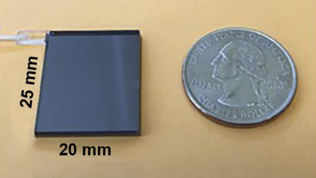
Figure 2. The first-generation eight-channel SDM-OCT chip measures 20 mm × 25 mm, approximately the size of a U.S. quarter. Adapted with permission from Reference 6.

Figure 3. A chip-based SDM-OCT image of an ex vivo porcine eyeball acquired in 1 s. This collage includes a single 2D cross-sectional B-scan showing eight images acquired simultaneously (a). 2D cross-sectional (b and c) and en face images (d) from the 3D data set. A 3D volumetric rendering of the porcine anterior chamber (e). Adapted with permission from Reference 6.
One of the main challenges in implementing PIC-based OCT is related to the practical wavelength in modern instruments. While OCT uses near-infrared light to image the eye, only specific wavelengths within this regime can effectively pass through the vitreous fluid and reach the retina. Ophthalmic OCT most commonly uses 850-nm and 1060-nm light, which have relatively low absorbance by the eye. On the contrary, current state-of-the-art PIC devices used for telecommunications generally use longer wavelengths, such as 1310 nm or 1550 nm, which are not suitable for retinal imaging.
Additionally, telecommunications typically operate in narrow wavelength ranges as opposed to the wide optical bandwidths (~100 nm) needed to achieve reasonable axial resolution in an OCT system. Therefore, a main hurdle in creating an effective PIC-OCT system for scientific and clinical purposes is optimizing all components and subsystems to support appropriate OCT wavelengths with broadband operation.
Since PIC fabrication uses many of the same processes as traditional CMOS and semiconductor manufacturing, it offers straightforward pathways to integrating many of the active components of an OCT system directly onto the chip. For example, materials such as germanium and indium gallium arsenide (InGaAs) can be deposited onto the PIC wafer, forming high-speed photodetectors, a crucial component in an OCT system. Crystal materials with nonlinear optical properties, such as lithium niobate, can be used to modulate optical path lengths, and laser gain materials can be integrated to provide optical amplification. In combination, these components can be used to construct a PIC-based laser source, thereby enabling the full consolidation of core OCT system functions onto a single chip-scale device.
Ongoing research has explored the development of photonic chip-based OCT featuring various architectures and varying levels of component integration. For example, a group at the Medical University of Vienna, led by Wolfgang Drexler, proposed a four-channel PIC-based swept-source OCT system with integrated photodetectors in 20207. In 2024, the same group also developed an integrated spectrometer for use in a spectral domain OCT system, which is slower than swept-source OCT8.
Another crucial challenge in the implementation of PIC-based OCT is the high cost and complexity of prototyping such devices, especially those that meet the performance requirements for OCT imaging. Fortunately, there is a rapidly growing number of opportunities for academic-commercial collaboration that will help foster a collaborative R&D ecosystem. For example, PIC foundries often offer a multi-project wafer (MPW) service, where multiple designs from different customers are aggregated and fabricated on a single wafer. Sharing the wafer space in this manner drastically reduces the upfront fabrication costs and reduces the barrier to entry for academic labs looking to order small quantities of devices for prototyping.
Even with MPW strategies, the complexity and subsequent cost of PIC device development rapidly increases with the addition of multiple advanced materials and optoelectronic components on the same chip. A fully integrated OCT system would potentially require a high-speed tunable laser, photodetectors, waveguides, and a handful of other solutions for electrical control and packaging. To address these challenges, the authors’ group has been awarded a grant from the Advanced Research Projects Agency for Health (ARPA-H) to develop the critical building blocks of a fully integrated PIC-based OCT system.
The prospect of a PIC-based OCT system is certainly exciting and holds great potential not only for ophthalmology, but for the OCT community at large. Although the current work is primarily focused on eye imaging, a generalized approach to constructing high-performance, miniaturized OCT systems could benefit a wide array of disciplines that use OCT, including cardiology, endoscopy, dermatology, dentistry, gynecology, and otolaryngology, among others. The field of integrated photonics for biomedical imaging is a highly active area of academic research, but its industrial development and commercialization are still in a relatively nascent state. Bridging this gap will require coordinated efforts between academia, industry, and clinical partners to translate lab-scale innovations into clinically viable tools.
Meet the authors
Aaron J. Adkins is a doctoral student in the Imaging Science Program at Washington University in St. Louis. He has a master’s degree in electrical engineering from Washington University and a bachelor’s degree in physics from Skidmore College; email: a.adkins@wustl.edu.
Senyue Hao is a doctoral student in the Department of Electrical and Systems Engineering at Washington University in St. Louis. He has a bachelor’s degree in computer science and engineering from the Chinese University of Hong Kong, Shenzhen; email: h.senyue@wustl.edu.
Chao Zhou is a professor in the Department of Biomedical Engineering at Washington University in St. Louis. He is a fellow in Optica, SPIE, and the American Heart Association, and a senior member of the Institute of Electrical and Electronics Engineers (IEEE). He received both his master’s degree and Ph.D. in physics from the University of Pennsylvania, and his bachelor’s degree in physics from Peking University; email: chaozhou@wustl.edu.
References
1. D.B. Rein et al. (2022). The economic burden of vision loss and blindness in the United States. Ophthalmology, Vol. 129, No. 4, pp. 369-378, www.doi.org/10.1016/j.ophtha.2021.09.010.
2. D. Huang et al. (1991). Optical coherence tomography. Science, Vol. 254, No. 5035, pp. 1178-1181.
3. Lasker Foundation (2023). OCT—for rapid detection of diseases of the retina, www.laskerfoundation.org/winners/optical-coherence-tomography.
4. C. Zhou et al. (2013). Space-division multiplexing optical coherence tomography. Opt Express, Vol. 21, No. 16, pp. 19219-19227, www.doi.org/10.1364/oe.21.019219.
5. J. Jerwick et al. (2020). Wide-field ophthalmic space-division multiplexing optical coherence tomography. Photon Res, Vol. 8, No. 4, pp. 539-547, www.doi.org/10.1364/prj.383034.
6. Y. Huang et al. (2017). Wide-field high-speed space-division multiplexing optical coherence tomography using an integrated photonic device. Biomed Opt Express, Vol. 8, No. 8, pp. 3856-3867.
7. S. Nevlacsil et al. (2020). Multi-channel swept source optical coherence tomography concept based on photonic integrated circuits. Opt Express, Vol. 28,
No. 22, pp. 32468-32482, www.doi.org/10.1364/oe.404588.
8. A. Agneter et al. (2024). CMOS optoelectronic spectrometer based on photonic integrated circuit for in vivo 3D optical coherence tomography. PhotoniX,
Vol. 5, No. 1, p. 31, www.doi.org/10.1186/s43074-024-00150-7.