A look at how photonics helps art be art, and at where the state of the art is headed.
Hank Hogan, Contributing Editor
Art may be for art’s sake, but that doesn’t mean that paintings and other works of art can’t benefit from photonic-based tools and equipment. For example, the challenge of preserving and restoring artwork from the past and present is made easier by techniques such as reflected infrared imaging and surface-enhanced Raman spectroscopy.
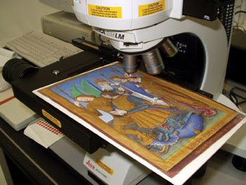
You cannot tell a book by its cover, but you can discover something about it by using spectroscopy. Here, a manuscript leaf by Jean Bourdichon, “The Portrait of Louis XII,” from “The Hours of Louis XII,” 1498 (J. Paul Getty Museum, MS 79a) is examined using noninvasive Raman microspectroscopy. With Raman spectroscopic analysis, investigators are characterizing the pigments used by Bourdichon as part of a larger study into the artist’s painting techniques and their relation to those of other 15th-century illuminators. Courtesy of Getty Conservation Institute.
Photonics also is helping adjust illumination to find the right balance between the light that is needed to see the art and damaging light that can shorten the lifetime of a painting, mural or other artwork.
And photonics may one day help solve today’s challenges. For instance, moving a fragile piece into a lab for analysis could harm it. The alternative, taking a sample, also does damage, although with modern techniques, the removed amount is often too small to be visible. The best solution may be performing analysis on the spot, which requires the development of portable instruments.
“A big tendency in conservation science that is going to be more so in the years to come is the use of non-destructive portable instruments,” noted Giacomo Chiari, chief scientist at Getty Conservation Institute in Los Angeles.
Friend and foe
At the institute, a team of 25 scientists uses many analytical imaging techniques, exploiting photons spanning the spectrum from infrared to visible to ultraviolet to x-rays. Measuring the interaction of photons of various wavelengths with an art object provides a wealth of information, thanks to such instruments as a portable x-ray fluorescence device from Bruker AXS of Madison, Wis., and a portable Raman spectrometer from DeltaNu Inc. of Laramie, Wyo. (now part of Intevac Corp. of Santa Clara, Calif.).
Sometimes that interaction leads to emission at a different wavelength, and ultraviolet fluorescence in particular can be revealing about the materials in the artwork, noted Chiari — provided care is taken. “It has to be done very well, with subtraction of all the parasitic light and with calibration. Then you get very good information on the distribution of very difficult to find, for example, organic material in a fresco.”
Armed with such information, conservators can determine the best way to restore a piece. Data on the material’s interaction with light also can serve another purpose. Museums often darken rooms to reduce light exposure and aging of the artwork while still allowing it to be viewable. A better solution might be to tailor the wavelength of the lighting by removing the strongly absorbed spectrum while preserving what people need for viewing. An added bonus of such an approach is that it could be more energy-efficient.
For all such benefits, there are also potential problems and real limitations with photon-based analytical techniques. The problems can arise from the use of a high-intensity source, such as the lasers found in some setups. Too much power will make what is supposed to be a nondestructive technique anything but. The limitations occur because most photon interactions are surface events. Hence, they cannot provide any information about the material deep within a sample.
One solution would be to use a confocal microscope or its x-ray fluorescence equivalent to optically section a sample and provide a layer-by-layer measurement. The latter might require technology that does not yet exist, such as a small x-ray tube that does not need cooling but that provides enough flux to allow rapid readings. “The idea to be able to in the future completely rely on nondestructive techniques, I think, for the moment, is still far away,” Chiari said.
Re-creating Homer
Francesca Casadio, a conservation scientist at the Art Institute of Chicago, is a member of a staff that is considerably smaller than that found at Getty Conservation Institute. Casadio is hard at work with paper conservator Kristi Dahm preparing for an exhibition of the master American watercolorist Winslow Homer, scheduled to open in February 2008 at the Chicago institute.
The many organic pigments used by the artist have faded over the years. The exhibition will use digital renderings to reconstruct the original images. Doing that requires determining exactly which pigments were used — a challenge because only minuscule particles are available for identification.
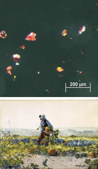
This micrograph (top) of pigment particles removed with a tungsten needle from the upper right corner of the sky in a watercolor painting (bottom) confirms that the work originally depicted an orange sunset, now lost because the red lake pigment faded. The pigment particles visible in the image have been analyzed and identified as chrome yellow, vermilion and an unidentified red lake. Techniques such as Raman spectroscopy and surface-enhanced Raman spectroscopy are used in the analysis of these tiny pigment particles. Photograph of the watercolor by Winslow Homer (American, 1863-1910), “For to Be a Farmer’s Boy,” 1887, a gift of Mrs. George T. Langhorne in memory of Edward Carson Waller (AIC 1963.760). Courtesy of the Art Institute of Chicago, conservation department.
For the most part, conventional Raman spectroscopy works for this task because the technique provides a chemical fingerprint using a particle as small as a micron. However, with particular red pigments, called red lakes, that were used by Homer in some paintings, that is not the case. “They are very fluorescent,” Casadio said. “The fluorescence swamps the Raman spectrum, and you don’t see the fingerprints.”
For that reason she has turned to surface-enhanced Raman spectroscopy, using a microscope from Horiba Jobin Yvon and substrate developed by Richard van Duyne at Northwestern University in Evanston, Ill. In surface-enhanced Raman spectroscopy, the normal Raman signal of a substance brought into contact with a nanostructured metallic substrate is amplified a millionfold or more, thanks to light-induced plasmons, or electron oscillations, in the metal surface. At the same time, the fluorescence gets quenched.
The problem has been getting repeatable and reproducible results, but the substrate developed at Northwestern eliminates this issue. Consisting of silica spheres overlaid with silver, the substrate provides the repeating structure needed by surface-enhanced Raman spectroscopy, and only one peak — that of silicon — arises from the substrate. Casadio noted that the spectra are extremely reproducible, and that only nanograms of material are needed for identification, although it is necessary to rid the sample of contaminants before applying the technique.
She is now working to identify the materials for the curators so that they will better know how to reproduce what has not been seen for a century or so. She cautioned, though, that these efforts will not result in perfection. “The reconstruction will be subjective because of course we can identify the pigment, but there are many nuances, shadings the artist could have applied,” she said.
Below the surface
People use different angles and vantage points to look at a painting. Henry Lie, director of the Straus Center for Conservation at the Harvard University Art Museums in Cambridge, Mass., likes to really get into art — as in below the surface. Although painted images capture the imagination, below the visible image are initial drawings and other elements. For example, a grid layout might signify that the design was actually taken from another painting.

There is more to a painting than meets the eye. Paint obscures any underdrawing in this detail (left) of “The Arrest of Christ,” circa 1520, from the Harvard University Art Museums. However, when viewed using surface-penetrating but substrate-reflected infrared, hidden features of the original drawing emerge (right). Courtesy of Henry Lie, Harvard University Art Museums.
One way to assess such images is through infrared reflectography, which exploits the fact that IR penetrates the surface pigments but is reflected by the prepared surface of the canvas or wood panel beneath. The technique has made significant contributions to the study of Northern European paintings.
Lie used a 256 × 256-pixel platinum silicide camera made by Inframetrics (since acquired by FLIR Systems Inc. of Boston) as well as a camera from Phase One A/S of Frederiksberg, Denmark.
For the most part, there is no need to go far into the infrared to do this. The Phase One camera, for instance, is sensitive only to 1.1 μm but yields good results when the paint is neither too thick nor too opaque. In other cases, a sensor with response out to 1.8 μm will work because most paints are transparent from 1.5 to 1.8 μm. Unfortunately, IR sensors are relatively small in terms of pixel count, and Lie noted that this presents a challenge when close-up details are desired.
“Because the paintings are sometimes large, almost no normal IR-type sensors are going to be adequate,” he said. “So you have to mosaic the images together.”
Capturing the IR image of a painting may require taking and stitching together 300 images. Mechanical setups have been built to make this possible without falling prey to parallax and other problems that would render an overlay useless. Lie would like to have an IR sensor that has more pixels and is part of a device offering good clarity and image quality. He acknowledged that getting an entire painting in a single shot probably will not be possible, but he would like to get the information he needs with fewer than 300 images.
Putting pieces together
Although no one has yet built that perfect IR imager for art, there continue to be technical advancements. Last year at the National Gallery in London, for example, researchers demonstrated a new camera, the scanning infrared imaging system (SIRIS), that is based on a 320 × 256-pixel indium gallium arsenide sensor array from Princeton, N.J.-based SUI, part of the Goodrich Corp. Through translation stages, the sensor is moved across the 5000 × 5000-pixel focal plane of the camera. A full image takes less than 20 minutes, faster than with sequential capture and mosaic.
Joseph Padfield, a conservation scientist at the National Gallery, was not part of the camera development team but is familiar with the device. He noted that, with better cameras, there might be a need to look for better light sources. Ideally, such a light source would be of sturdy construction and would have a stable, diffuse output with a good spectral match to the sensor. “It would also be good if the lights did not give off a lot of thermal IR —wavelengths higher than the camera sensitivity — so that the object being examined would not heat up,” he said.
On Padfield’s wish list are devices with more speed, better resolution, higher bit depth, greater color accuracy and spectral control, and greater cost-effectiveness.
As more and more digital imaging data becomes available, image processing may become in the near future an important part of art conservation and preservation. For example, Padfield pointed to an application of polynomial texture mapping Hewlett-Packard software with raking light, an examination done with a light source at a very low angle. The software, originally developed for three-dimensional rendering, accurately simulated the effect of having the light anywhere, revealing new details.
Padfield also talked about a viewer that allows two images to be overlaid, as long as they have been correctly prepared and registered. Controls enable the user to zoom in on an image and to view images separately as well as in combination. This device could be used, for example, to examine visible and x-ray images of the same painting. He said that art conservators are beginning to use similar viewers within the gallery to disseminate large images where appropriate.
Caring for the young
At the New York-based Museum of Modern Art (MoMA), chief conservator James Coddington deals with issues of age, but he does not have to fret over pieces that are centuries old. Instead, the artwork at MoMA is only decades old or, in some cases, years old.
“We’re caring for works that are very young,” he noted. “They’re comparatively pristine.”
That immaculate condition makes it possible to capture information about the object before pigments dim and materials dull. For that purpose, Coddington has turned to multispectral imaging, using a commercial infrared camera and narrow-band filters to capture the reflectance spectrum of a work of art from 1.1 to 2.0 μm. He also has done something similar in the visible, using a custom-built system developed by Roy Berns of Rochester Institute of Technology. The setup consisted of a high-end red-green-blue (RBG) sensor array with a filter wheel to get six peaks, instead of the normal three, in the visible.
The unspoiled state of young art does not last forever, and stopping the degradation can be difficult. In particular, people need light to see an object, but light also can attack artwork.
In estimating such effects, Coddington has made use of another piece of photonic gear, a microfading tester developed by Paul M. Whitmore of Carnegie Mellon University. This device sends light from a high-intensity source down a fiber optic cable so that a point on the object is exposed. The result is captured by a spectrophotometer, which accurately measures the reflectance. From a brief exposure of a small area, a linear rate of change can be established, and guidelines about display lighting conditions, such as intensity and time, can be set.
Although such techniques find the weakest link, frequently the news is not bad. The unexpected robustness brings artwork out of the dark, Coddington reported. “We can feel confident about displaying it a little bit longer.”